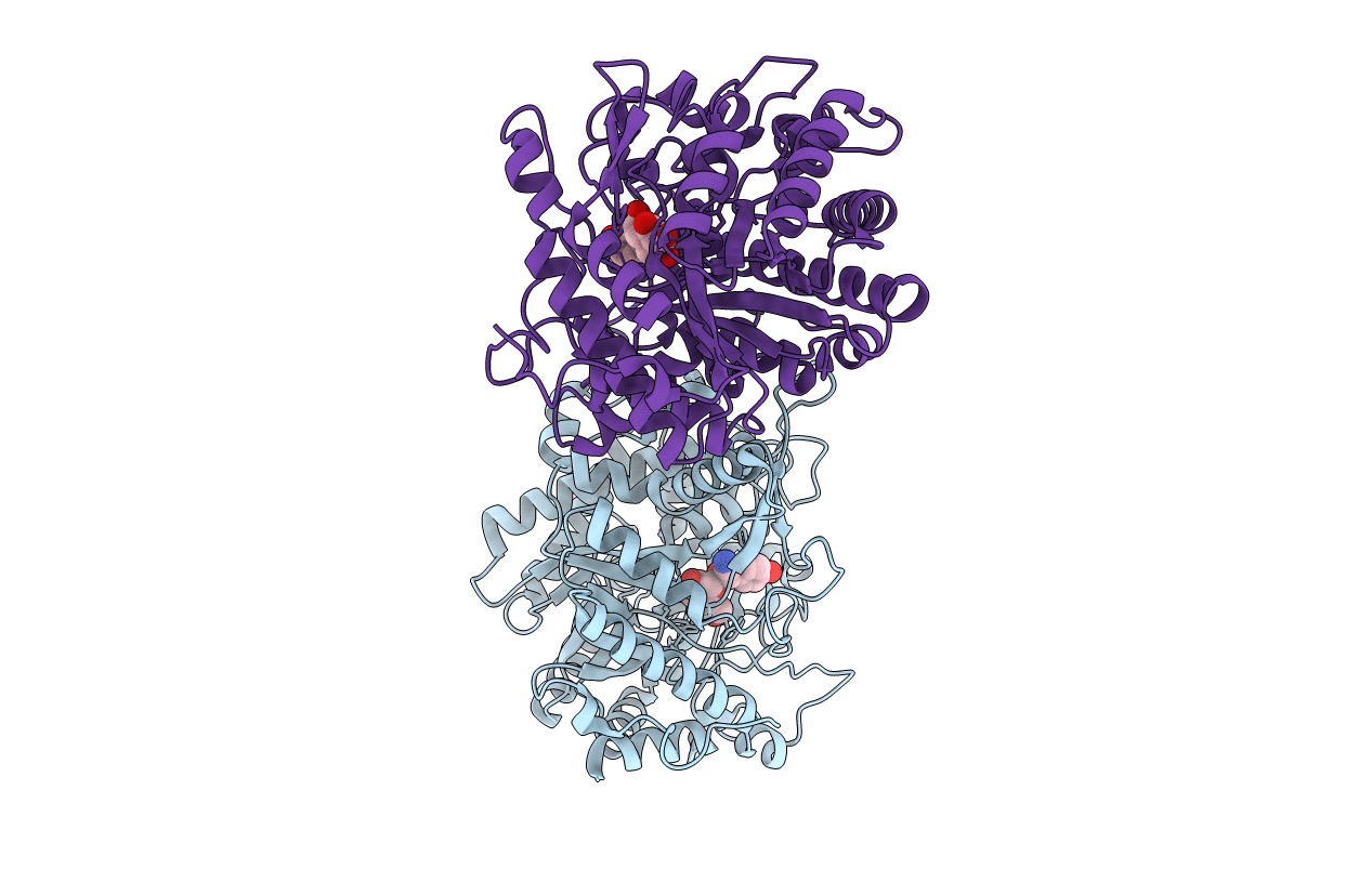
1E55 image
Entry Detail
PDB ID:
1E55
Keywords:
Title:
Crystal structure of the inactive mutant Monocot (Maize ZMGlu1) beta-glucosidase ZMGluE191D in complex with the competitive inhibitor dhurrin
Biological Source:
Source Organism:
Host Organism:
PDB Version:
Deposition Date:
2000-07-18
Release Date:
2000-12-11
Method Details:
Experimental Method:
Resolution:
2.00 Å
R-Value Free:
0.23
R-Value Work:
0.19
R-Value Observed:
0.19
Space Group:
P 21 21 21
Macromolecular Entities
Polymer Type:polypeptide(L)
Description:BETA-GLUCOSIDASE
Mutations:YES
Chain IDs:A, B
Chain Length:512
Number of Molecules:2
Biological Source:ZEA MAYS
Primary Citation
The mechanism of substrate (aglycone) specificity in beta-glucosidases is revealed by crystal structures of mutant maize beta-glucosidase-DIMBOA, -DIMBOAGlc, and -dhurrin complexes.
Proc. Natl. Acad. Sci. U.S.A. 97 13555 13560 (2000)
PMID: 11106394 DOI: 10.1073/pnas.97.25.13555

Abstact

The mechanism and the site of substrate (i.e., aglycone) recognition and specificity were investigated in maize beta-glucosidase (Glu1) by x-ray crystallography by using crystals of a catalytically inactive mutant (Glu1E191D) in complex with the natural substrate 2-O-beta-d-glucopyranosyl-4-hydroxy-7-methoxy-1,4-benzoxazin-3-one (DIMBOAGlc), the free aglycone DIMBOA, and competitive inhibitor para-hydroxy-S-mandelonitrile beta-glucoside (dhurrin). The structures of these complexes and of the free enzyme were solved at 2.1-, 2.1-, 2.0-, and 2.2-A resolution, respectively. The structural data from the complexes allowed us to visualize an intact substrate, free aglycone, or a competitive inhibitor in the slot-like active site of a beta-glucosidase. These data show that the aglycone moiety of the substrate is sandwiched between W378 on one side and F198, F205, and F466 on the other. Thus, specific conformations of these four hydrophobic amino acids and the shape of the aglycone-binding site they form determine aglycone recognition and substrate specificity in Glu1. In addition to these four residues, A467 interacts with the 7-methoxy group of DIMBOA. All residues but W378 are variable among beta-glucosidases that differ in substrate specificity, supporting the conclusion that these sites are the basis of aglycone recognition and binding (i.e., substrate specificity) in beta-glucosidases. The data also provide a plausible explanation for the competitive binding of dhurrin to maize beta-glucosidases with high affinity without being hydrolyzed.

Legend

Protein

Chemical

Disease

Primary Citation of related structures