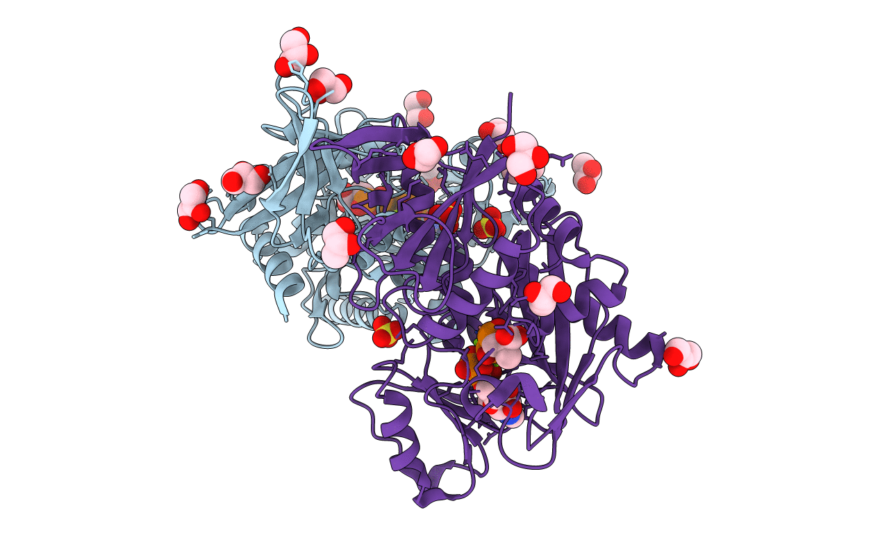
1E4E image
Deposition Date 2000-07-03
Release Date 2001-06-28
Last Version Date 2024-10-09
Entry Detail
PDB ID:
1E4E
Keywords:
Title:
D-alanyl-D-lacate ligase
Biological Source:
Source Organism:
Host Organism:
Method Details:
Experimental Method:
Resolution:
2.50 Å
R-Value Free:
0.25
R-Value Work:
0.18
Space Group:
C 2 2 21
Macromolecular Entities
Polymer Type:polypeptide(L)
Molecule:VANCOMYCIN/TEICOPLANIN A-TYPE RESISTANCE PROTEIN VANA
Gene (Uniprot):vanA
Chain IDs:A
Chain Length:343
Number of Molecules:1
Biological Source:ENTEROCOCCUS FAECIUM
Polymer Type:polypeptide(L)
Molecule:VANCOMYCIN/TEICOPLANIN A-TYPE RESISTANCE PROTEIN VANA
Gene (Uniprot):vanA
Chain IDs:B
Chain Length:343
Number of Molecules:1
Biological Source:ENTEROCOCCUS FAECIUM
Primary Citation
The molecular basis of vancomycin resistance in clinically relevant Enterococci: crystal structure of D-alanyl-D-lactate ligase (VanA).
Proc. Natl. Acad. Sci. U.S.A. 97 8921 8925 (2000)
PMID: 10908650 DOI: 10.1073/pnas.150116497

Abstact

d-alanine-d-lactate ligase from Enterococcus faecium BM4147 is directly responsible for the biosynthesis of alternate cell-wall precursors in bacteria, which are resistant to the glycopeptide antibiotic vancomycin. The crystal structure has been determined with data extending to 2.5-A resolution. This structure shows that the active site has unexpected interactions and is distinct from previous models for d-alanyl-d-lactate ligase mechanistic studies. It appears that the preference of the enzyme for lactate as a ligand over d-alanine could be mediated by electrostatic effects and/or a hydrogen-bonding network, which principally involve His-244. The structure of d-alanyl-d-lactate ligase provides a revised interpretation of the molecular events that lead to vancomycin resistance.

Legend

Protein

Chemical

Disease

Primary Citation of related structures