
Deposition Date
2000-06-26
Release Date
2001-06-21
Last Version Date
2024-05-08
Entry Detail
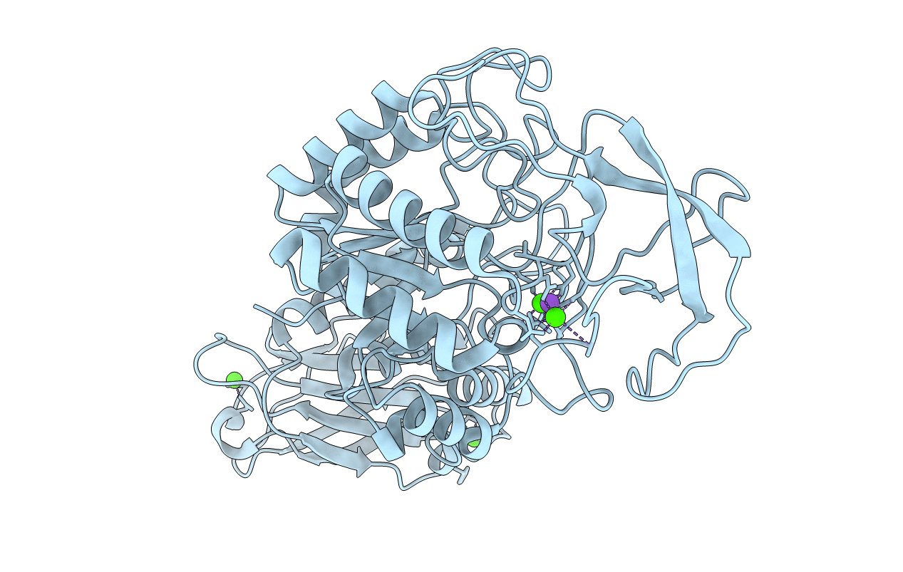
PDB ID:
1E3X
Keywords:
Title:
Native structure of chimaeric amylase from B. amyloliquefaciens and B. licheniformis at 1.92A
Biological Source:
Source Organism(s):
BACILLUS AMYLOLIQUEFACIENS (Taxon ID: 1390)
Expression System(s):
Method Details:
Experimental Method:
Resolution:
1.90 Å
R-Value Free:
0.20
R-Value Work:
0.14
Space Group:
C 2 2 21
