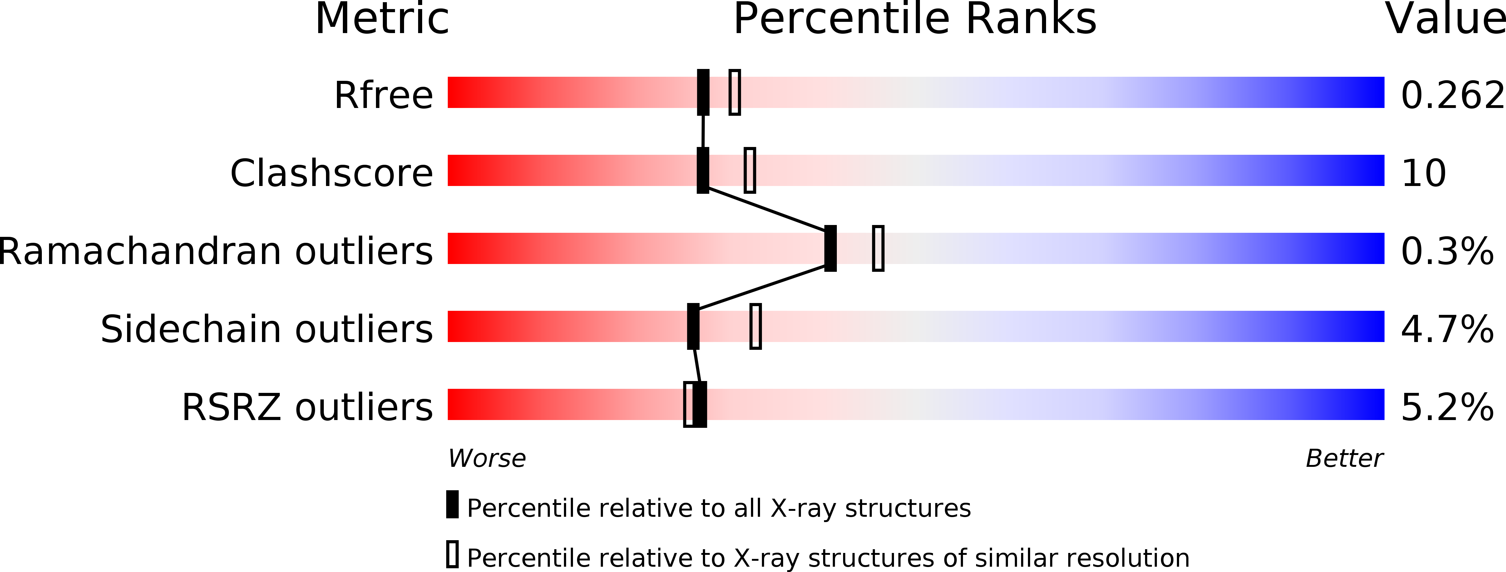
1E3M image
Deposition Date 2000-06-19
Release Date 2000-11-01
Last Version Date 2024-10-23
Entry Detail
PDB ID:
1E3M
Keywords:
Title:
The crystal structure of E. coli MutS binding to DNA with a G:T mismatch
Biological Source:
Source Organism:
ESCHERICHIA COLI (Taxon ID: 562)
Host Organism:
Method Details:
Experimental Method:
Resolution:
2.20 Å
R-Value Free:
0.26
R-Value Work:
0.22
R-Value Observed:
0.22
Space Group:
P 21 21 21
Macromolecular Entities
Polymer Type:polypeptide(L)
Molecule:DNA MISMATCH REPAIR PROTEIN MUTS
Gene (Uniprot):mutS
Chain IDs:A, B
Chain Length:800
Number of Molecules:2
Biological Source:ESCHERICHIA COLI
Polymer Type:polydeoxyribonucleotide
Molecule:5'-D(*AP*GP*CP*TP*GP*CP*CP*AP*GP*GP*CP*AP*CP*CP*AP* GP*TP*GP*TP*CP*AP*GP*CP*GP*TP*CP*CP*TP*AP*T)-3'
Chain IDs:C (auth: E)
Chain Length:30
Number of Molecules:1
Biological Source:ESCHERICHIA COLI
Polymer Type:polydeoxyribonucleotide
Molecule:5'-D(*AP*TP*AP*GP*GP*AP*CP*GP*CP*TP*GP*AP*CP*AP*CP* TP*GP*GP*TP*GP*CP*TP*TP*GP*GP*CP*AP*GP*CP*T)-3'
Chain IDs:D (auth: F)
Chain Length:30
Number of Molecules:1
Biological Source:ESCHERICHIA COLI
Modified Residue
Compound ID Chain ID Parent Comp ID Details 2D Image
MSE A MET SELENOMETHIONINE
Primary Citation
The Crystal Structure of DNA Mismatch Repair Protein Muts Binding to a G X T Mismatch
Nature 407 711 ? (2000)
PMID: 11048711 DOI: 10.1038/35037523

Abstact

DNA mismatch repair ensures genomic integrity on DNA replication. Recognition of a DNA mismatch by a dimeric MutS protein initiates a cascade of reactions and results in repair of the newly synthesized strand; however, details of the molecular mechanism remain controversial. Here we present the crystal structure at 2.2 A of MutS from Escherichia coli bound to a G x T mismatch. The two MutS monomers have different conformations and form a heterodimer at the structural level. Only one monomer recognizes the mismatch specifically and has ADP bound. Mismatch recognition occurs by extensive minor groove interactions causing unusual base pairing and kinking of the DNA. Nonspecific major groove DNA-binding domains from both monomers embrace the DNA in a clamp-like structure. The interleaved nucleotide-binding sites are located far from the DNA. Mutations in human MutS alpha (MSH2/MSH6) that lead to hereditary predisposition for cancer, such as hereditary non-polyposis colorectal cancer, can be mapped to this crystal structure.

Legend

Protein

Chemical

Disease

Primary Citation of related structures