1E3H image
Deposition Date 2000-06-15
Release Date 2000-11-05
Last Version Date 2024-10-23
Entry Detail
PDB ID:
1E3H
Title:
SeMet derivative of Streptomyces antibioticus PNPase/GPSI enzyme
Biological Source:
Source Organism:
Host Organism:
Method Details:
Experimental Method:
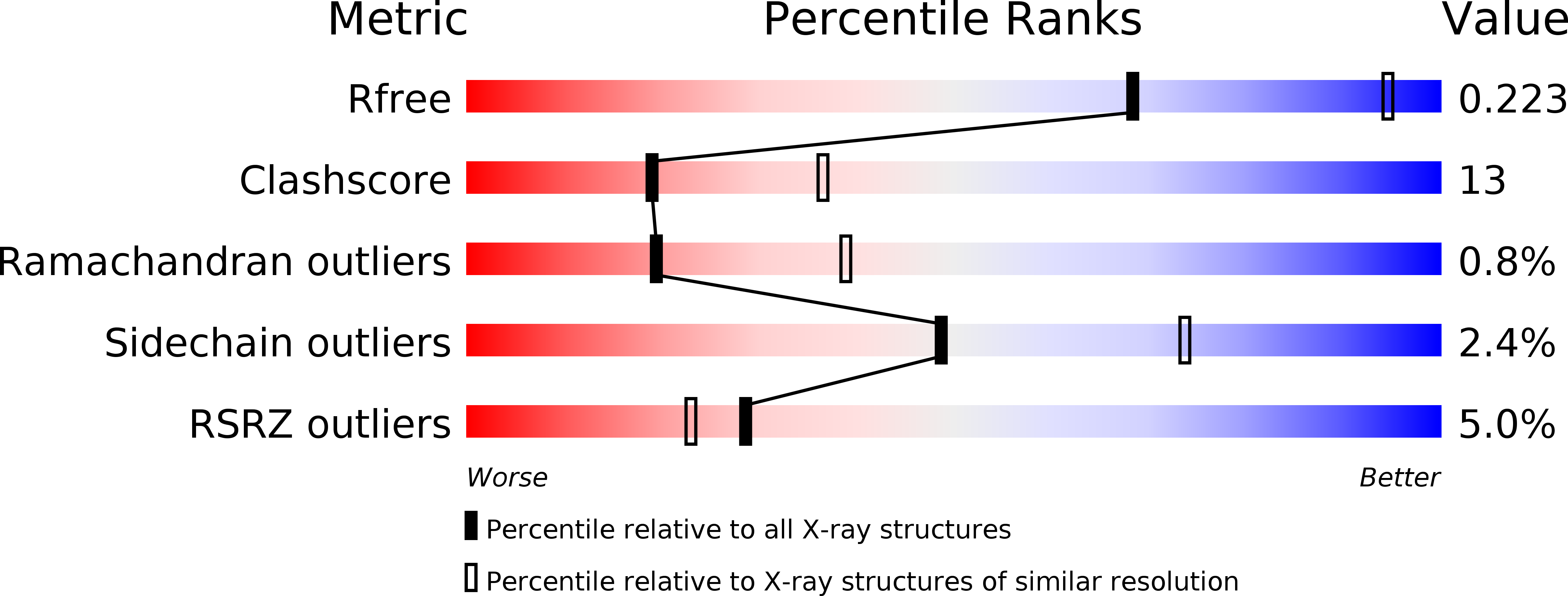
Resolution:
2.60 Å
R-Value Free:
0.22
R-Value Work:
0.20
R-Value Observed:
0.20
Space Group:
H 3 2
Macromolecular Entities
Structures with similar UniProt ID
Protein Blast
Polymer Type:polypeptide(L)
Molecule:GUANOSINE PENTAPHOSPHATE SYNTHETASE
Gene (Uniprot):pnp
Chain IDs:A
Chain Length:757
Number of Molecules:1
Biological Source:STREPTOMYCES ANTIBIOTICUS
Modified Residue
Compound ID Chain ID Parent Comp ID Details 2D Image
MSE A MET SELENOMETHIONINE
Ligand Molecules
Primary Citation
A Duplicated Fold is the Structural Basis for Polynucleotide Phosphorylase Catalytic Activity, Processivity, and Regulation
Structure 8 1215 ? (2000)
PMID: 11080643 DOI: 10.1016/S0969-2126(00)00521-9

Abstact

BACKGROUND: Polynucleotide phosphorylase (PNPase) is a polyribonucleotide nucleotidyl transferase (E.C.2.7.7.8) that degrades mRNA in prokaryotes. Streptomyces antibioticus PNPase also assays as a guanosine 3'-diphosphate 5'-triphosphate (pppGpp) synthetase (E.C.2.7.6.5). It may function to coordinate changes in mRNA lifetimes with pppGpp levels during the Streptomyces lifecycle. RESULTS: The structure of S. antibioticus PNPase without bound RNA but with the phosphate analog tungstate bound at the PNPase catalytic sites was determined by X-ray crystallography and shows a trimeric multidomain protein with a central channel. The structural core has a novel duplicated architecture formed by association of two homologous domains. The tungstate derivative structure reveals the PNPase active site in the second of these core domains. Structure-based sequence analysis suggests that the pppGpp synthetase active site is located in the first core domain. CONCLUSIONS: This is the first structure of a PNPase and shows the structural basis for the trimer assembly, the arrangement of accessory RNA binding domains, and the likely catalytic residues of the PNPase active site. A possible function of the trimer channel is as a contribution to both the processivity of degradation and the regulation of PNPase action by RNA structural elements.

Legend

Protein

Chemical

Disease

Primary Citation of related structures