1E2B image
Deposition Date 1996-11-15
Release Date 1997-07-23
Last Version Date 2024-05-22
Entry Detail
PDB ID:
1E2B
Keywords:
Title:
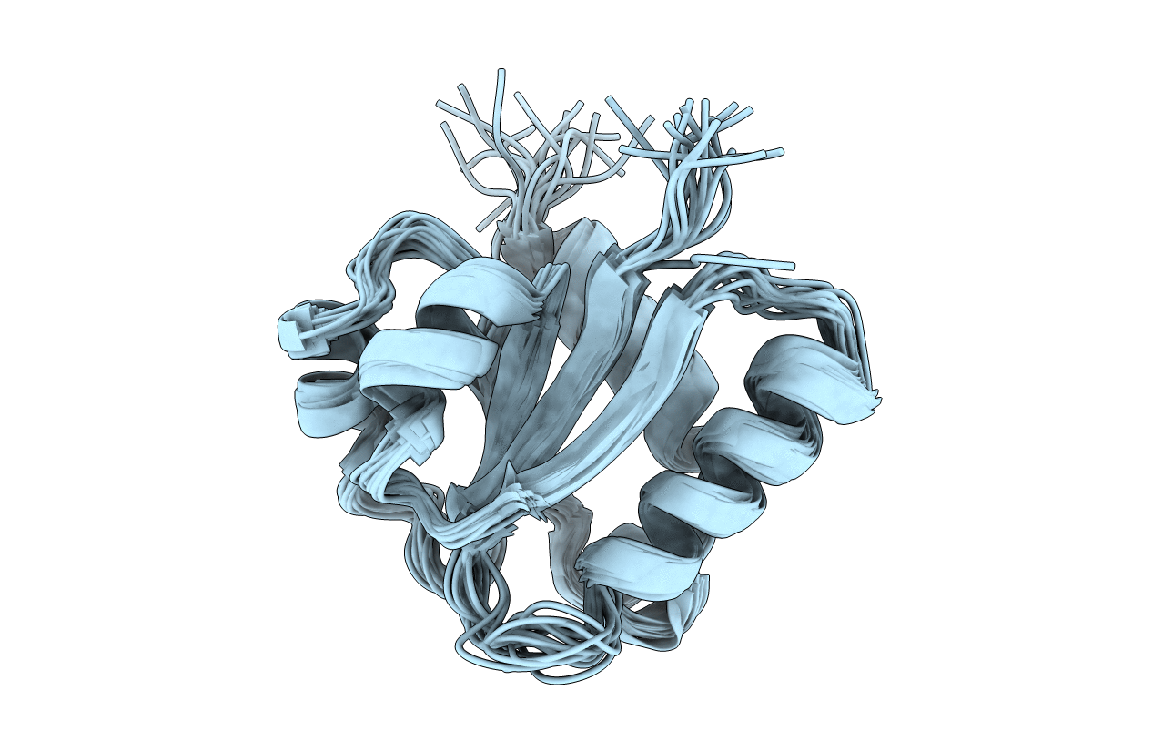
NMR STRUCTURE OF THE C10S MUTANT OF ENZYME IIB CELLOBIOSE OF THE PHOSPHOENOL-PYRUVATE DEPENDENT PHOSPHOTRANSFERASE SYSTEM OF ESCHERICHIA COLI, 17 STRUCTURES
Biological Source:
Source Organism:
Method Details:
Experimental Method:
Conformers Calculated:
32
Conformers Submitted:
17
Selection Criteria:
TARGET FUNCTION, NUMBER OF VIOLATIONS
Macromolecular Entities
Polymer Type:polypeptide(L)
Molecule:ENZYME IIB-CELLOBIOSE
Gene (Uniprot):chbB
Mutations:C10S
Chain IDs:A
Chain Length:106
Number of Molecules:1
Biological Source:Escherichia coli
Ligand Molecules
Primary Citation
The NMR side-chain assignments and solution structure of enzyme IIBcellobiose of the phosphoenolpyruvate-dependent phosphotransferase system of Escherichia coli.
Protein Sci. 6 304 314 (1997)
PMID: 9041631

Abstact

The assignment of the side-chain NMR resonances and the determination of the three-dimensional solution structure of the C10S mutant of enzyme IIBcellobiose (IIBcel) of the phosphoenolpyruvate-dependent phosphotransferase system of Escherichia coli are presented. The side-chain resonances were assigned nearly completely using a variety of mostly heteronuclear NMR experiments, including HCCH-TOCSY, HCCH-COSY, and COCCH-TOCSY experiments as well as CBCACOHA, CBCA(CO)NH, and HBHA(CBCA)(CO)NH experiments. In order to obtain the three-dimensional structure, NOE data were collected from 15N-NOESY-HSQC, 13C-HSQC-NOESY, and 2D NOE experiments. The distance restraints derived from these NOE data were used in distance geometry calculations followed by molecular dynamics and simulated annealing protocols. In an iterative procedure, additional NOE assignments were derived from the calculated structures and new structures were calculated. The final set of structures, calculated with approximately 2000 unambiguous and ambiguous distance restraints, has an rms deviation of 1.1 A on C alpha atoms. IIBcel consists of a four stranded parallel beta-sheet, in the order 2134. The sheet is flanked with two and three alpha-helices on either side. Residue 10, a cysteine in the wild-type enzyme, which is phosphorylated during the catalytic cycle, is located at the end of the first beta-strand. A loop that is proposed to be involved in the binding of the phosphoryl-group follows the cysteine. The loop appears to be disordered in the unphosphorylated state.

Legend

Protein

Chemical

Disease

Primary Citation of related structures