Deposition Date
2000-02-25
Release Date
2000-08-16
Last Version Date
2024-10-23
Entry Detail
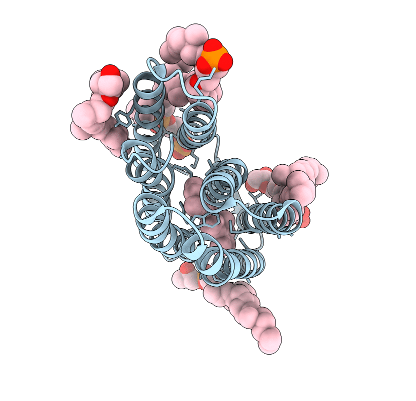
PDB ID:
1DZE
Keywords:
Title:
Structure of the M Intermediate of Bacteriorhodopsin trapped at 100K
Biological Source:
Source Organism(s):
HALOBACTERIUM SALINARIUM (Taxon ID: 2242)
Method Details:
Experimental Method:
Resolution:
2.50 Å
R-Value Free:
0.28
R-Value Work:
0.25
R-Value Observed:
0.25
Space Group:
P 6 2 2