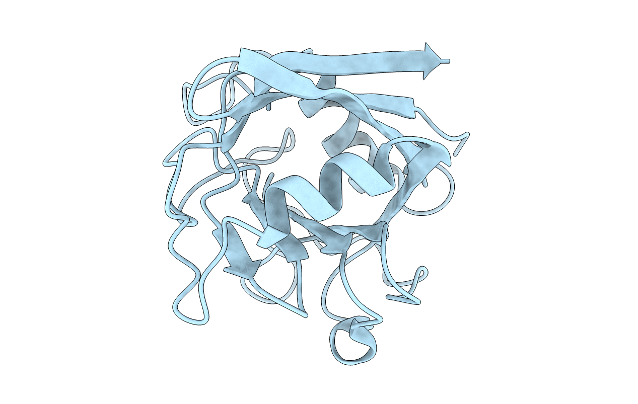
1DYW image
Deposition Date 2000-02-10
Release Date 2000-06-22
Last Version Date 2023-12-06
Entry Detail
PDB ID:
1DYW
Title:
Biochemical and structural characterization of a divergent loop cyclophilin from Caenorhabditis elegans
Biological Source:
Source Organism:
Host Organism:
Method Details:
Experimental Method:
Resolution:
1.80 Å
R-Value Free:
0.28
R-Value Observed:
0.21
Space Group:
P 41 21 2
Macromolecular Entities
Structures with similar UniProt ID
Protein Blast
Polymer Type:polypeptide(L)
Molecule:CYCLOPHILIN 3
Gene (Uniprot):cyn-3
Chain IDs:A
Chain Length:173
Number of Molecules:1
Biological Source:CAENORHABDITIS ELEGANS
Primary Citation
Biochemical and Structural Characterization of a Divergent Loop Cyclophilin from Caenorhabditis Elegans
J.Biol.Chem. 274 34877 ? (1999)
PMID: 10574961 DOI: 10.1074/JBC.274.49.34877

Abstact

Cyclophilin 3 (CYP-3) is one of the most abundantly expressed cyclophilin isoforms in the free living nematode Caenorhabditis elegans. The detailed post-embryonic expression pattern of the cyp-3 transcript is unusual, peaking during early larval development. The spatial expression pattern was examined via reporter gene analysis demonstrating that the cyp-3 transcript is exclusively expressed in the single anterior excretory cell. Recombinant cyclophilin 3 has been purified, crystallized and solved to a resolution of 1.8 A. The peptidyl-prolyl isomerase activity of CYP-3 has been characterized against the substrate N-succinyl-Ala-Ala-Pro-Phe-p-nitroanilide, and gives a k(cat)/K(m) value of 2.4 x 10(6) M(-1) s(-1). The immunosuppressive drug cyclosporin A binds and inhibits CYP-3 with an IC(50) value of 16 nM, comparable with the range of values found for human cyclophilin A. The x-ray structure shows that the overall fold and active site geometry is similar to other cyclophilin structures. There are however a number of distinctive features, and we use this structure and amino acid sequence alignment analysis to identify a subgroup of "divergent-loop cyclophilins". This subgroup has a number of uniquely conserved features: an additional loop between residues 48 and 54 (KSGKPLH); two cysteine residues (Cys(40) and Cys(168)) that are in close proximity but remain in the unoxidized form, and two other conserved residues, His(54) and Glu(83). We suggest that these features are functionally important for the role played by this class of cyclophilins during cellular responses to stress caused by changes in the redox environment or by up-regulation of cellular activity. This study represents a detailed biological, biochemical, and structural characterization of a single cyclophilin isoform in the model organism Caenorhabditis elegans.

Legend

Protein

Chemical

Disease

Primary Citation of related structures