Deposition Date
1994-12-21
Release Date
1995-02-27
Last Version Date
2024-02-07
Entry Detail
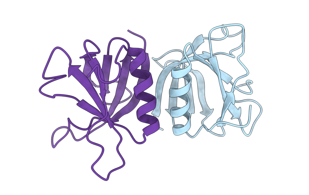
PDB ID:
1DYN
Keywords:
Title:
CRYSTAL STRUCTURE AT 2.2 ANGSTROMS RESOLUTION OF THE PLECKSTRIN HOMOLOGY DOMAIN FROM HUMAN DYNAMIN
Biological Source:
Source Organism(s):
Homo sapiens (Taxon ID: 9606)
Method Details:
Experimental Method:
Resolution:
2.20 Å
R-Value Free:
0.32
R-Value Work:
0.20
R-Value Observed:
0.20
Space Group:
I 4 2 2