Deposition Date
2000-01-31
Release Date
2001-01-28
Last Version Date
2024-11-20
Entry Detail
PDB ID:
1DY9
Keywords:
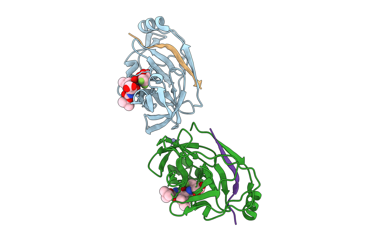
Title:
Inhibition of the Hepatitis C Virus NS3/4A Protease. The Crystal Structures of Two Protease-Inhibitor Complexes (inhibitor I)
Biological Source:
Source Organism(s):
HEPATITIS C VIRUS (ISOLATE TAIWAN) (Taxon ID: 31645)
Expression System(s):
Method Details:
Experimental Method:
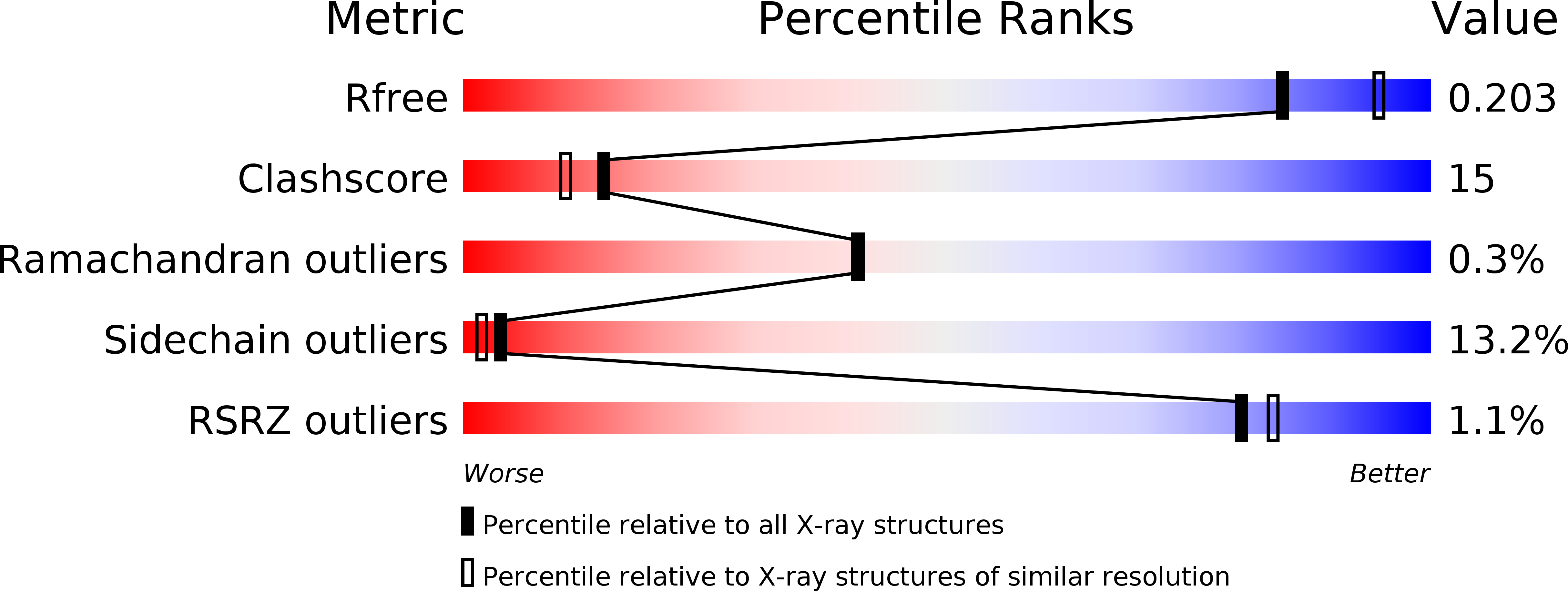
Resolution:
2.10 Å
R-Value Free:
0.27
R-Value Work:
0.21
R-Value Observed:
0.20
Space Group:
P 61