1DY7 image
Deposition Date 2000-01-28
Release Date 2000-09-24
Last Version Date 2024-11-06
Entry Detail
PDB ID:
1DY7
Keywords:
Title:
Cytochrome cd1 Nitrite Reductase, CO complex
Biological Source:
Source Organism(s):
Method Details:
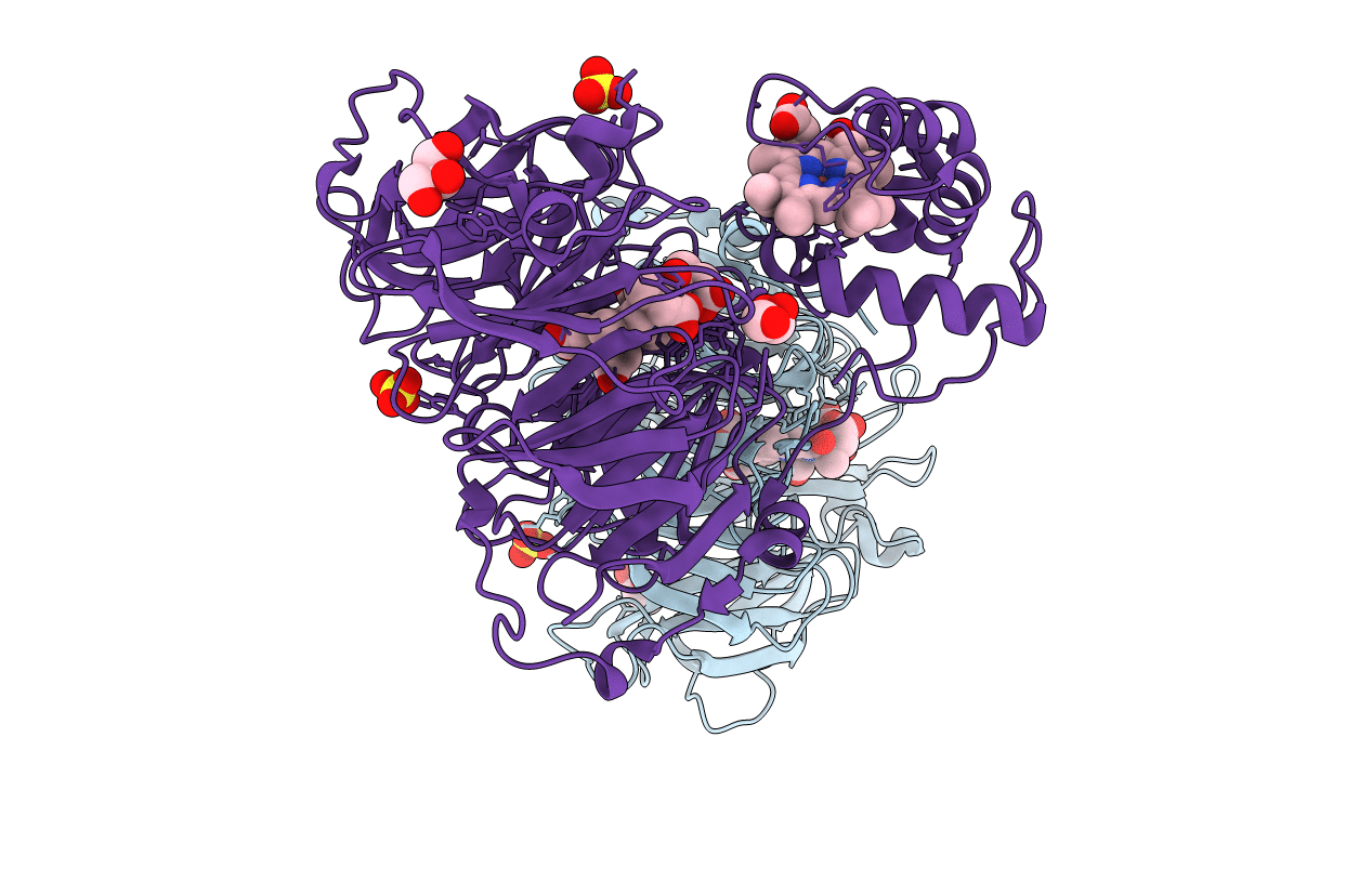
Experimental Method:
Resolution:
1.60 Å
R-Value Free:
0.19
R-Value Work:
0.17
Space Group:
P 1 21 1
Macromolecular Entities
Structures with similar UniProt ID
Protein Blast
Polymer Type:polypeptide(L)
Molecule:NITRITE REDUCTASE
Gene (Uniprot):nirS
Chain IDs:A, B
Chain Length:567
Number of Molecules:2
Biological Source:PARACOCCUS PANTOTROPHUS
Primary Citation
Proton-Coupled Structural Changes Upon Binding of Carbon Monoxide to Cytochrome Cd(1): A Combined Flash Photolysis and X-Ray Crystallography Study
Biochemistry 39 10967 ? (2000)
PMID: 10998233 DOI: 10.1021/BI000179Q

Abstact

We have investigated dynamic events after flash photolysis of CO from reduced cytochrome cd(1) nitrite reductase (NiR) from Paracoccus pantotrophus (formerly Thiosphaera pantotropha). Upon pulsed illumination of the cytochrome cd(1)-CO complex, at 460 nm, a rapid (<50 ns) absorbance change, attributed to dissociation of CO, was observed. This was followed by a biphasic rearrangement with rate constants of 1.7 x 10(4) and 2.5 x 10(3) s(-1) at pH 8.0. Both parts of the biphasic rearrangement phases displayed the same kinetic difference spectrum in the region of 400-660 nm. The slower of the two processes was accompanied by proton uptake from solution (0.5 proton per active site at pH 7.5-8.5). After photodissociation, the CO ligand recombined at a rate of 12 s(-1) (at 1 mM CO and pH 8.0), accompanied by proton release. The crystal structure of reduced cytochrome cd(1) in complex with CO was determined to a resolution of 1.57 A. The structure shows that CO binds to the iron of the d(1) heme in the active site. The ligation of the c heme is unchanged in the complex. A comparison of the structures of the reduced, unligated NiR and the NiR-CO complex indicates changes in the puckering of the d(1) heme as well as rearrangements in the hydrogen-bonding network and solvent organization in the substrate binding pocket at the d(1) heme. Since the CO ligand binds to heme d(1) and there are structural changes in the d(1) pocket upon CO binding, it is likely that the proton uptake or release observed after flash-induced CO dissociation is due to changes of the protonation state of groups in the active site. Such proton-coupled structural changes associated with ligand binding are likely to affect the redox potential of heme d(1) and may regulate the internal electron transfer from heme c to heme d(1).

Legend

Protein

Chemical

Disease

Primary Citation of related structures
Feedback Form
Name
Email
Institute
Feedback