Deposition Date
1999-12-07
Release Date
2000-05-16
Last Version Date
2024-10-23
Entry Detail
PDB ID:
1DWK
Keywords:
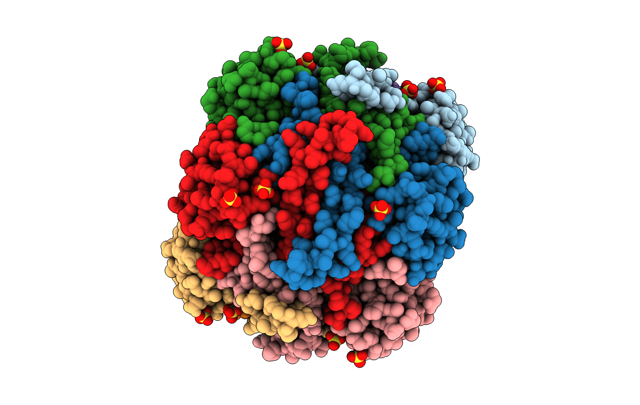
Title:
STRUCTURE OF CYANASE WITH THE DI-ANION OXALATE BOUND AT THE ENZYME ACTIVE SITE
Biological Source:
Source Organism(s):
ESCHERICHIA COLI (Taxon ID: 562)
Expression System(s):
Method Details:
Experimental Method:
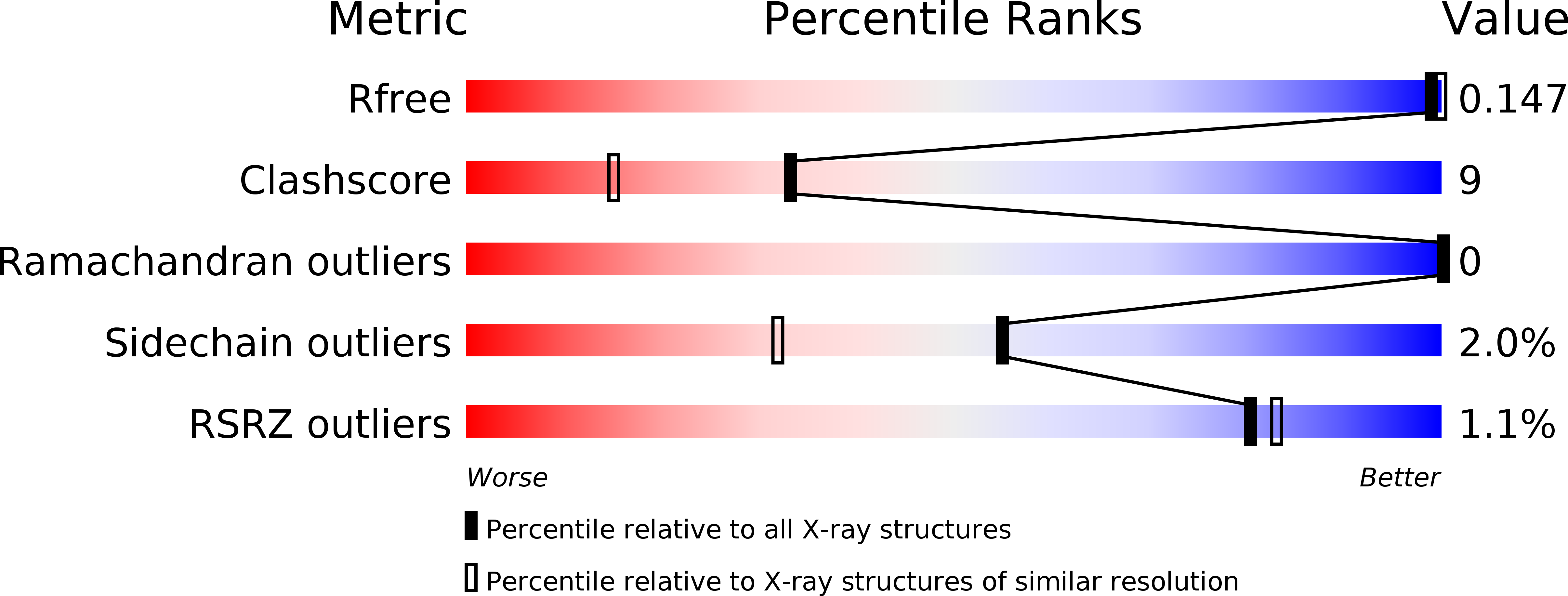
Resolution:
1.65 Å
R-Value Free:
0.18
R-Value Work:
0.14
R-Value Observed:
0.13
Space Group:
P 1