1DW9 image
Deposition Date 1999-12-03
Release Date 2000-05-16
Last Version Date 2024-11-13
Entry Detail
PDB ID:
1DW9
Keywords:
Title:
Structure of cyanase reveals that a novel dimeric and decameric arrangement of subunits is required for formation of the enzyme active site
Biological Source:
Source Organism:
ESCHERICHIA COLI (Taxon ID: 562)
Host Organism:
Method Details:
Experimental Method:
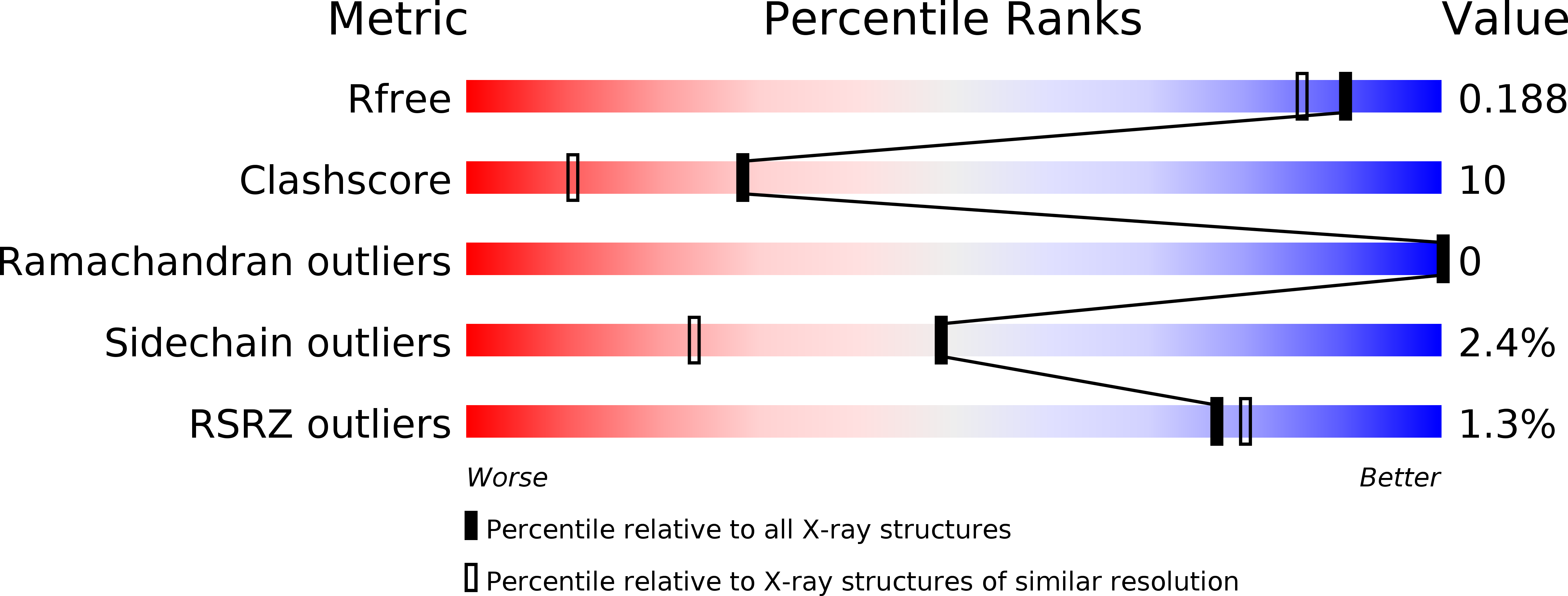
Resolution:
1.65 Å
R-Value Free:
0.18
R-Value Work:
0.15
Space Group:
P 1
Macromolecular Entities
Structures with similar UniProt ID
Protein Blast
Polymer Type:polypeptide(L)
Molecule:CYANATE LYASE
Gene (Uniprot):cynS
Chain IDs:A, B, C, D, E, F, G, H, I, J
Chain Length:156
Number of Molecules:10
Biological Source:ESCHERICHIA COLI
Modified Residue
Compound ID Chain ID Parent Comp ID Details 2D Image
MSE A MET SELENOMETHIONINE
Primary Citation
Structure of Cyanase Reveals that a Novel Dimeric and Decameric Arrangement of Subunits is Required for Formation of the Enzyme Active Site
Structure 8 505 ? (2000)
PMID: 10801492 DOI: 10.1016/S0969-2126(00)00134-9

Abstact

BACKGROUND: Cyanase is an enzyme found in bacteria and plants that catalyzes the reaction of cyanate with bicarbonate to produce ammonia and carbon dioxide. In Escherichia coli, cyanase is induced from the cyn operon in response to extracellular cyanate. The enzyme is functionally active as a homodecamer of 17 kDa subunits, and displays half-site binding of substrates or substrate analogs. The enzyme shows no significant amino acid sequence homology with other proteins. RESULTS: We have determined the crystal structure of cyanase at 1.65 A resolution using the multiwavelength anomalous diffraction (MAD) method. Cyanase crystals are triclinic and contain one homodecamer in the asymmetric unit. Selenomethionine-labeled protein offers 40 selenium atoms for use in phasing. Structures of cyanase with bound chloride or oxalate anions, inhibitors of the enzyme, allowed identification of the active site. CONCLUSIONS: The cyanase monomer is composed of two domains. The N-terminal domain shows structural similarity to the DNA-binding alpha-helix bundle motif. The C-terminal domain has an 'open fold' with no structural homology to other proteins. The subunits of cyanase are arranged in a novel manner both at the dimer and decamer level. The dimer structure reveals the C-terminal domains to be intertwined, and the decamer is formed by a pentamer of these dimers. The active site of the enzyme is located between dimers and is comprised of residues from four adjacent subunits of the homodecamer. The structural data allow a conceivable reaction mechanism to be proposed.

Legend

Protein

Chemical

Disease

Primary Citation of related structures
Feedback Form
Name
Email
Institute
Feedback