1DVO image
Entry Detail
PDB ID:
1DVO
Keywords:
Title:
THE X-RAY CRYSTAL STRUCTURE OF FINO, A REPRESSOR OF BACTERIAL CONJUGATION
Biological Source:
Source Organism:
Host Organism:
PDB Version:
Deposition Date:
2000-01-21
Release Date:
2000-07-19
Method Details:
Experimental Method:
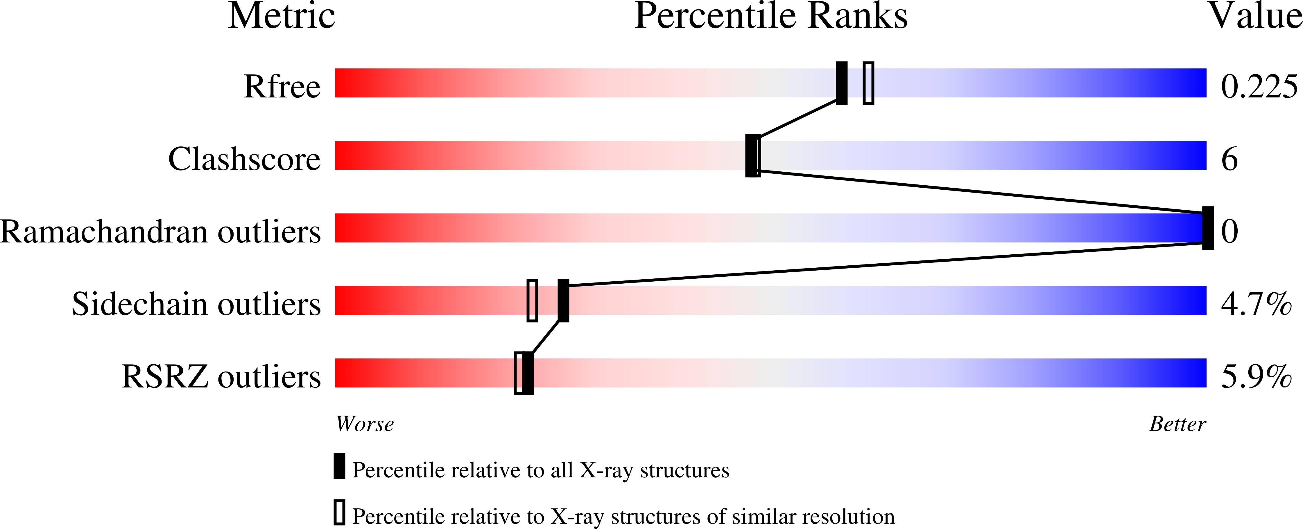
Resolution:
2.00 Å
R-Value Free:
0.22
R-Value Work:
0.19
Space Group:
P 21 21 21
Macromolecular Entities
Polymer Type:polypeptide(L)
Description:FERTILITY INHIBITION PROTEIN O
Mutations:L124M
Chain IDs:A
Chain Length:152
Number of Molecules:1
Biological Source:Escherichia coli
Primary Citation
Crystal structure of the bacterial conjugation repressor finO.
Nat.Struct.Biol. 7 565 569 (2000)
PMID: 10876242 DOI: 10.1038/76790

Abstact

The conjugative transfer of F-like plasmids is repressed by FinO, an RNA binding protein. FinO interacts with the F-plasmid encoded traJ mRNA and its antisense RNA, FinP, stabilizing FinP against endonucleolytic degradation and facilitating sense-antisense RNA recognition. Here we present the 2.0 A resolution X-ray crystal structure of FinO, lacking its flexible N-terminal extension. FinO adopts a novel, elongated, largely helical conformation. An N-terminal region, previously shown to contact RNA, forms a positively charged alpha-helix (helix 1) that protrudes 45 A from the central core of FinO. A C-terminal region of FinO that is implicated in RNA interactions also extends out from the central body of the protein, adopting a helical conformation and packing against the base of the N-terminal helix. A highly positively charged patch on the surface of the FinO core may present another RNA binding surface. The results of an in vitro RNA duplexing assay demonstrate that the flexible N-terminal region of FinO plays a key role in FinP-traJ RNA recognition, and supports our proposal that this region and the N-terminus of helix 1 interact with and stabilize paired, complementary RNA loops in a kissing complex.

Legend

Protein

Chemical

Disease

Primary Citation of related structures