Deposition Date
2000-01-20
Release Date
2000-04-12
Last Version Date
2024-10-30
Entry Detail
PDB ID:
1DVG
Keywords:
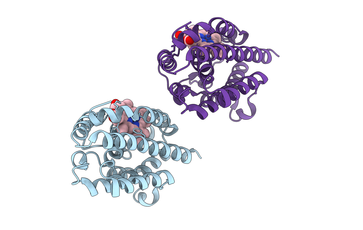
Title:
CRYSTAL STRUCTURE OF RAT HEME OXYGENASE-1 IN COMPLEX WITH HEME; SELELENO-METHIONINE DERIVATIVE, MUTATED AT M51T,M93L,M155L,M191L.
Biological Source:
Source Organism(s):
Rattus norvegicus (Taxon ID: 10116)
Expression System(s):
Method Details:
Experimental Method:
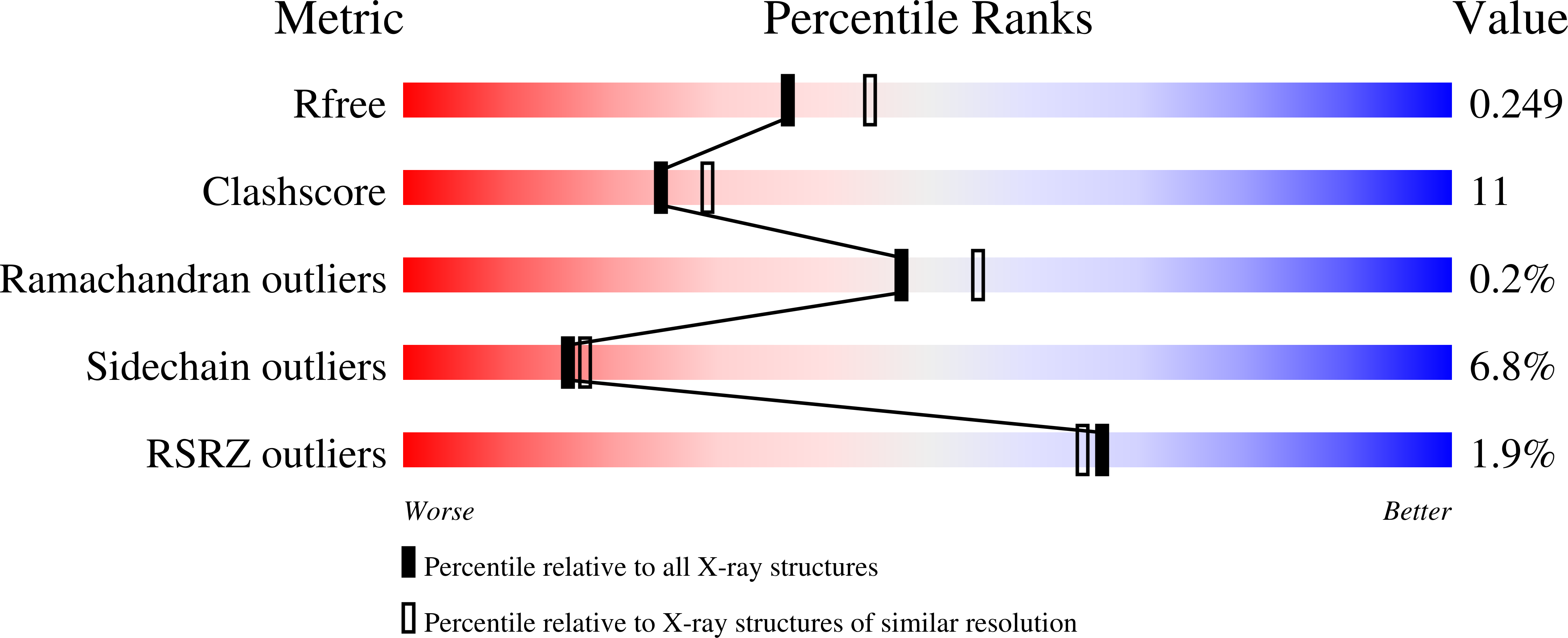
Resolution:
2.20 Å
R-Value Free:
0.25
R-Value Work:
0.21
Space Group:
P 43