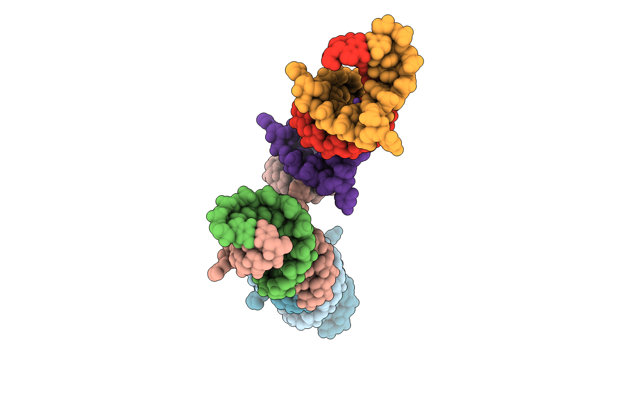
1DUQ image
Deposition Date 2000-01-18
Release Date 2000-05-16
Last Version Date 2024-02-07
Entry Detail
PDB ID:
1DUQ
Keywords:
Title:
CRYSTAL STRUCTURE OF THE REV BINDING ELEMENT OF HIV-1
Biological Source:
Source Organism:
(Taxon ID: )
Method Details:
Experimental Method:
Resolution:
2.10 Å
R-Value Free:
0.26
R-Value Work:
0.21
R-Value Observed:
0.21
Space Group:
P 1
Macromolecular Entities
Polymer Type:polyribonucleotide
Molecule:THE REV BINDING ELEMENT
Chain IDs:A, C, E, G
Chain Length:12
Number of Molecules:4
Biological Source:
Ligand Molecules
Primary Citation
The crystal structure of the Rev binding element of HIV-1 reveals novel base pairing and conformational variability.
Proc.Natl.Acad.Sci.USA 97 5107 5112 (2000)
PMID: 10792052 DOI: 10.1073/pnas.090588197

Abstact

The crystal and molecular structure of an RNA duplex corresponding to the high affinity Rev protein binding element (RBE) has been determined at 2.1-A resolution. Four unique duplexes are present in the crystal, comprising two structural variants. In each duplex, the RNA double helix consists of an annealed 12-mer and 14-mer that form an asymmetric internal loop consisting of G-G and G-A noncanonical base pairs and a flipped-out uridine. The 12-mer strand has an A-form conformation, whereas the 14-mer strand is distorted to accommodate the bulges and noncanonical base pairing. In contrast to the NMR model of the unbound RBE, an asymmetric G-G pair with N2-N7 and N1-O6 hydrogen bonding, is formed in each helix. The G-A base pairing agrees with the NMR structure in one structural variant, but forms a novel water-mediated pair in the other. A backbone flip and reorientation of the G-G base pair is required to assume the RBE conformation present in the NMR model of the complex between the RBE and the Rev peptide.

Legend

Protein

Chemical

Disease

Primary Citation of related structures