1DT9 image
Entry Detail
PDB ID:
1DT9
Keywords:
Title:
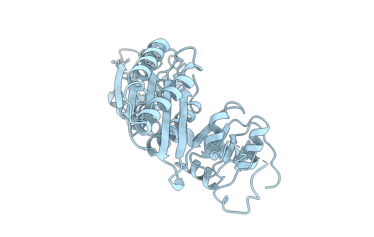
THE CRYSTAL STRUCTURE OF HUMAN EUKARYOTIC RELEASE FACTOR ERF1-MECHANISM OF STOP CODON RECOGNITION AND PEPTIDYL-TRNA HYDROLYSIS
Biological Source:
Source Organism:
PDB Version:
Deposition Date:
2000-01-12
Release Date:
2000-02-02
Method Details:
Experimental Method:
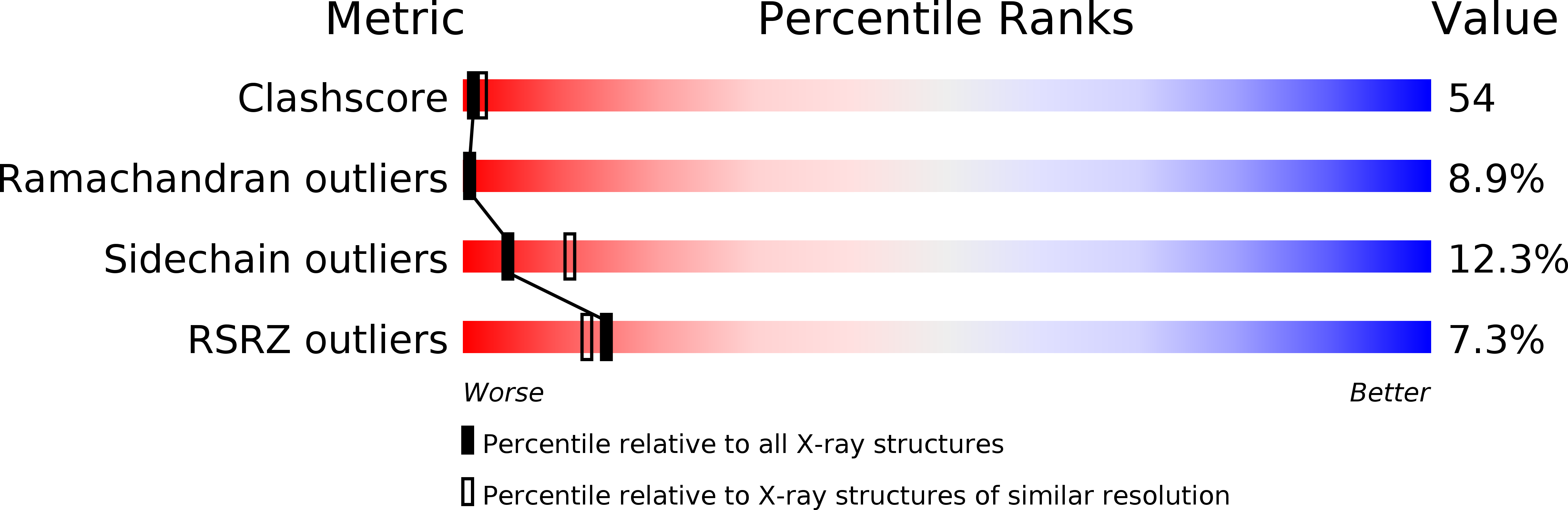
Resolution:
2.70 Å
R-Value Free:
0.31
R-Value Work:
0.24
Space Group:
P 43 21 2
Macromolecular Entities
Polymer Type:polypeptide(L)
Description:PROTEIN (EUKARYOTIC PEPTIDE CHAIN RELEASE FACTOR SUBUNIT 1)
Chain IDs:A
Chain Length:437
Number of Molecules:1
Biological Source:Homo sapiens
Primary Citation
The crystal structure of human eukaryotic release factor eRF1--mechanism of stop codon recognition and peptidyl-tRNA hydrolysis.
Cell(Cambridge,Mass.) 100 311 321 (2000)
PMID: 10676813 DOI: 10.1016/S0092-8674(00)80667-4

Abstact

The release factor eRF1 terminates protein biosynthesis by recognizing stop codons at the A site of the ribosome and stimulating peptidyl-tRNA bond hydrolysis at the peptidyl transferase center. The crystal structure of human eRF1 to 2.8 A resolution, combined with mutagenesis analyses of the universal GGQ motif, reveals the molecular mechanism of release factor activity. The overall shape and dimensions of eRF1 resemble a tRNA molecule with domains 1, 2, and 3 of eRF1 corresponding to the anticodon loop, aminoacyl acceptor stem, and T stem of a tRNA molecule, respectively. The position of the essential GGQ motif at an exposed tip of domain 2 suggests that the Gln residue coordinates a water molecule to mediate the hydrolytic activity at the peptidyl transferase center. A conserved groove on domain 1, 80 A from the GGQ motif, is proposed to form the codon recognition site.

Legend

Protein

Chemical

Disease

Primary Citation of related structures