Deposition Date
1993-05-24
Release Date
1994-01-31
Last Version Date
2024-10-09
Entry Detail
PDB ID:
1DSB
Keywords:
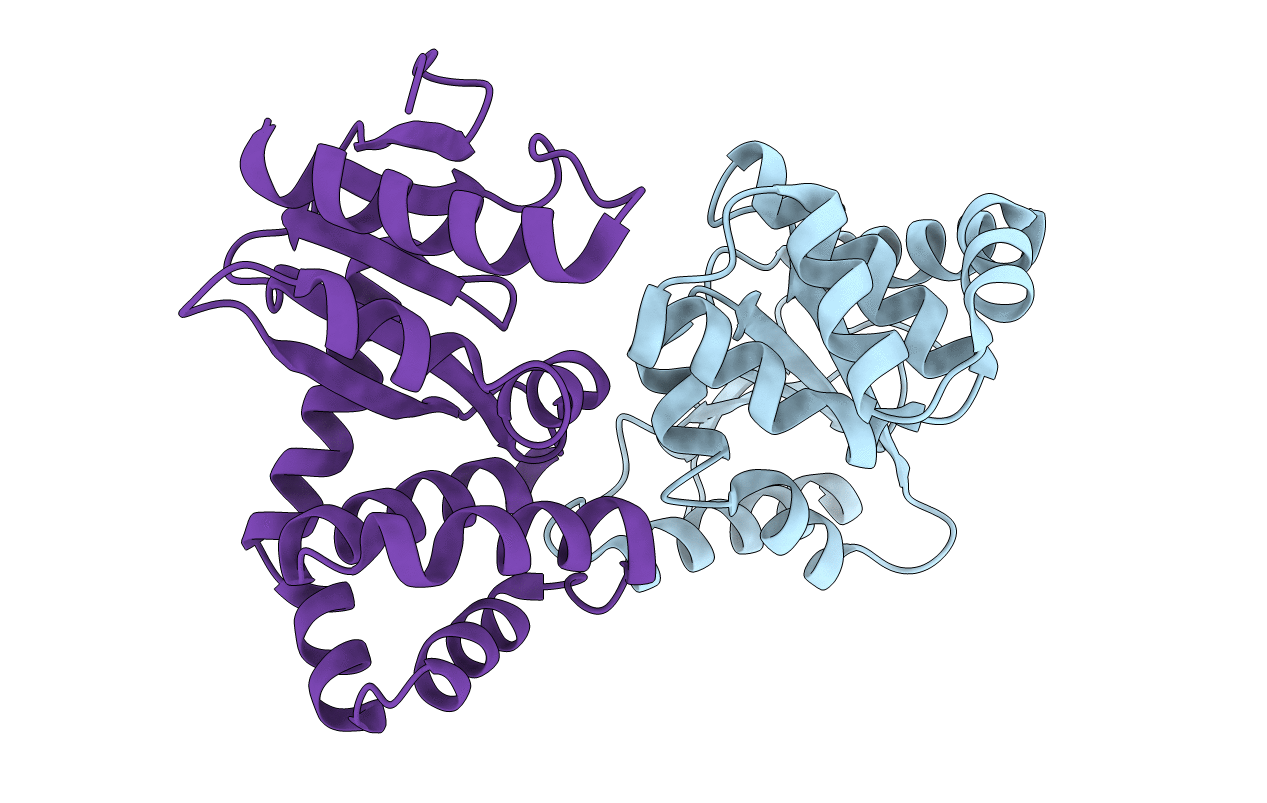
Title:
CRYSTAL STRUCTURE OF THE DSBA PROTEIN REQUIRED FOR DISULPHIDE BOND FORMATION IN VIVO
Biological Source:
Source Organism(s):
Escherichia coli (Taxon ID: 562)
Method Details:
Experimental Method:
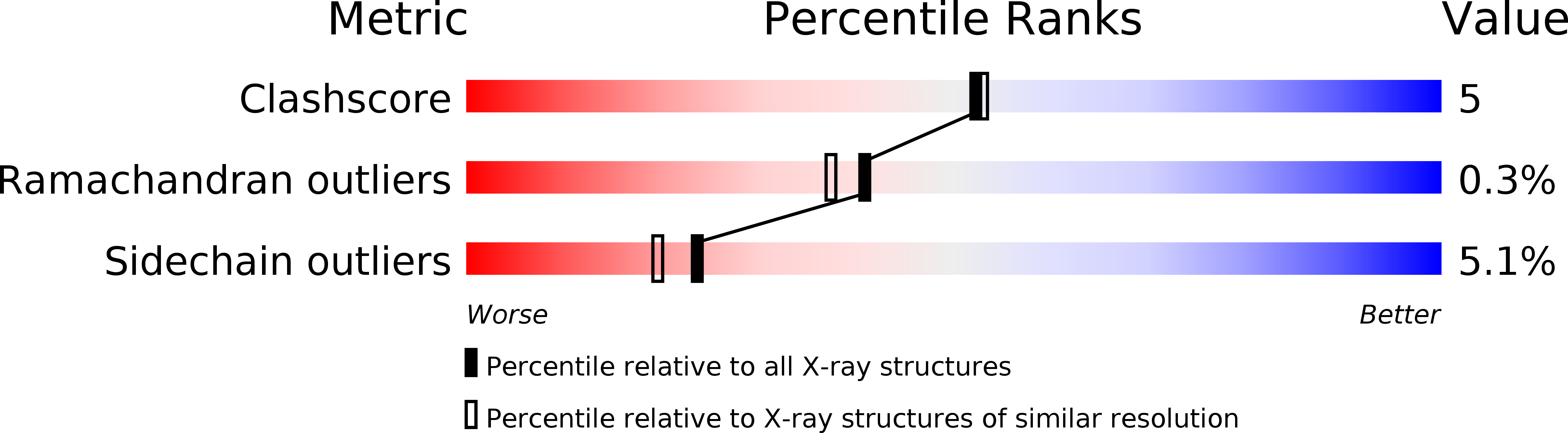
Resolution:
2.00 Å
R-Value Work:
0.16
R-Value Observed:
0.16
Space Group:
C 1 2 1