1DQX image
Deposition Date 2000-01-05
Release Date 2000-03-20
Last Version Date 2024-02-07
Entry Detail
PDB ID:
1DQX
Keywords:
Title:
CRYSTAL STRUCTURE OF OROTIDINE 5'-PHOSPHATE DECARBOXYLASE COMPLEXED TO 6-HYDROXYURIDINE 5'-PHOSPHATE (BMP)
Biological Source:
Source Organism:
Host Organism:
Method Details:
Experimental Method:
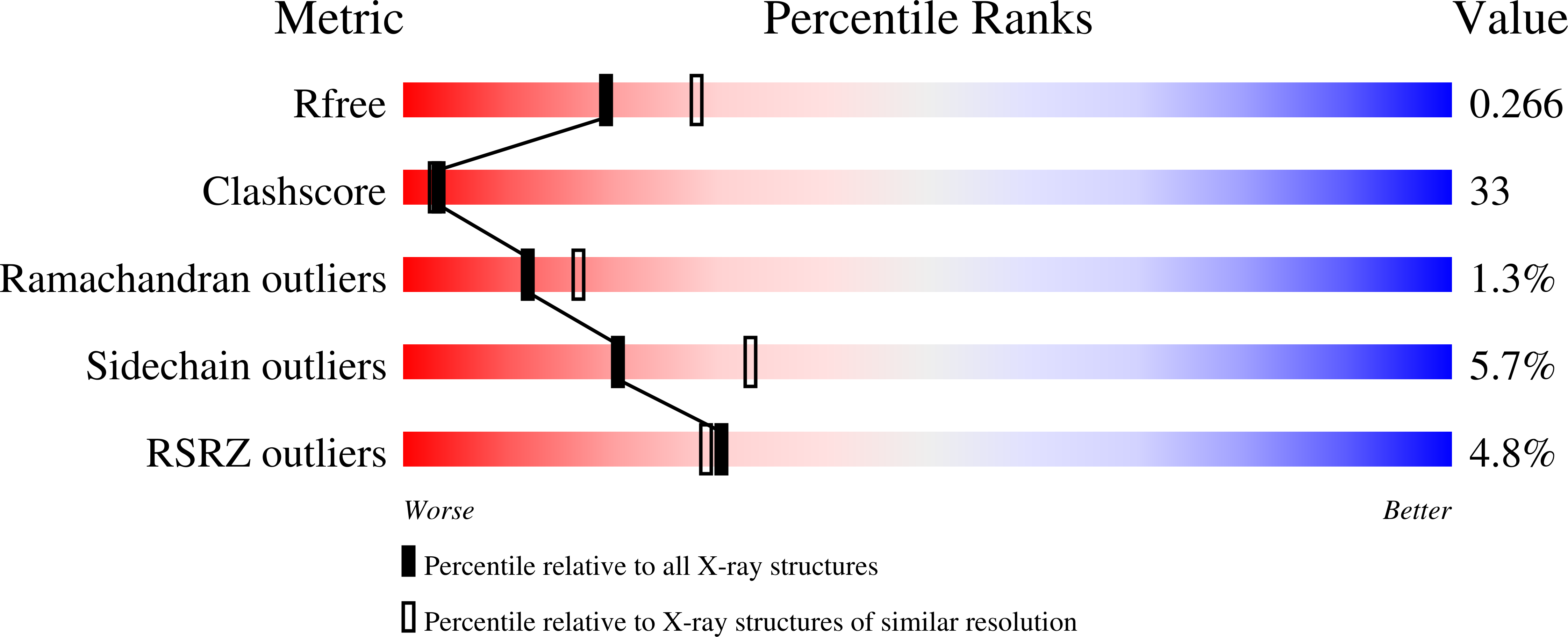
Resolution:
2.40 Å
R-Value Free:
0.25
R-Value Work:
0.21
R-Value Observed:
0.21
Space Group:
P 1 21 1
Macromolecular Entities
Polymer Type:polypeptide(L)
Molecule:OROTIDINE 5'-PHOSPHATE DECARBOXYLASE
Gene (Uniprot):URA3
Mutations:S2H, N267D
Chain IDs:A, B, C, D
Chain Length:267
Number of Molecules:4
Biological Source:Saccharomyces cerevisiae
Ligand Molecules
Primary Citation
Anatomy of a proficient enzyme: the structure of orotidine 5'-monophosphate decarboxylase in the presence and absence of a potential transition state analog.
Proc.Natl.Acad.Sci.USA 97 2011 2016 (2000)
PMID: 10681417 DOI: 10.1073/pnas.030409797

Abstact

Orotidine 5'-phosphate decarboxylase produces the largest rate enhancement that has been reported for any enzyme. The crystal structure of the recombinant Saccharomyces cerevisiae enzyme has been determined in the absence and presence of the proposed transition state analog 6-hydroxyuridine 5'-phosphate, at a resolution of 2.1 A and 2.4 A, respectively. Orotidine 5'-phosphate decarboxylase folds as a TIM-barrel with the ligand binding site near the open end of the barrel. The binding of 6-hydroxyuridine 5'-phosphate is accompanied by protein loop movements that envelop the ligand almost completely, forming numerous favorable interactions with the phosphoryl group, the ribofuranosyl group, and the pyrimidine ring. Lysine-93 appears to be anchored in such a way as to optimize electrostatic interactions with developing negative charge at C-6 of the pyrimidine ring, and to donate the proton that replaces the carboxylate group at C-6 of the product. In addition, H-bonds from the active site to O-2 and O-4 help to delocalize negative charge in the transition state. Interactions between the enzyme and the phosphoribosyl group anchor the pyrimidine within the active site, helping to explain the phosphoribosyl group's remarkably large contribution to catalysis despite its distance from the site of decarboxylation.

Legend

Protein

Chemical

Disease

Primary Citation of related structures