1DQV image
Entry Detail
PDB ID:
1DQV
Title:
CRYSTAL STRUCTURE OF SYNAPTOTAGMIN III C2A/C2B
Biological Source:
Source Organism:
Host Organism:
PDB Version:
Deposition Date:
2000-01-05
Release Date:
2000-01-19
Method Details:
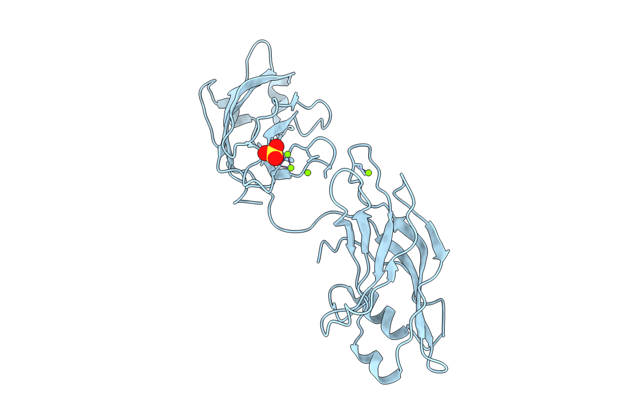
Experimental Method:
Resolution:
3.20 Å
R-Value Free:
0.34
R-Value Work:
0.29
R-Value Observed:
0.29
Space Group:
P 62 2 2
Macromolecular Entities
Polymer Type:polypeptide(L)
Description:SYNAPTOTAGMIN III
Chain IDs:A
Chain Length:296
Number of Molecules:1
Biological Source:Rattus rattus
Primary Citation
Crystal structure of the cytosolic C2A-C2B domains of synaptotagmin III. Implications for Ca(+2)-independent snare complex interaction.
J.Cell Biol. 147 589 598 (1999)
PMID: 10545502 DOI: 10.1083/jcb.147.3.589

Abstact

Synaptotagmins are synaptic vesicle-associated, phospholipid-binding proteins most commonly associated with Ca(+2)-dependent exocytotic and Ca(+2)- independent endocytotic events. Synaptotagmin III is a 63.2-kD member of the synaptotagmin homology group; one of its characteristic properties is the ability to bind divalent cations and accessory proteins promiscuously. In the cytosolic portion of this protein, a flexible seven-amino acid linker joins two homologous C2 domains. The C2A domain binds to phospholipid membranes and other accessory proteins in a divalent cation-dependent fashion. The C2B domain promotes binding to other C2B domains, as well as accessory proteins independent of divalent cations. The 3.2 A crystal structure of synaptotagmin III, residues 295-566, which includes the C2A and C2B domains, exhibits differences in the shape of the Ca(+2)-binding pocket, the electrostatic surface potential, and the stoichiometry of bound divalent cations for the two domains. These observations may explain the disparate binding properties of the two domains. The C2A and the C2B domains do not interact; synaptotagmin, therefore, covalently links two independent C2 domains, each with potentially different binding partners. A model of synaptotagmin's involvement in Ca(+2)-dependent regulation of membrane fusion through its interaction with the SNARE complex is presented.

Legend

Protein

Chemical

Disease

Primary Citation of related structures