Deposition Date
2000-01-05
Release Date
2000-05-10
Last Version Date
2024-02-07
Entry Detail
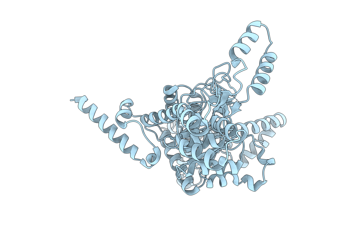
PDB ID:
1DQU
Keywords:
Title:
CRYSTAL STRUCTURE OF THE ISOCITRATE LYASE FROM ASPERGILLUS NIDULANS
Biological Source:
Source Organism:
Emericella nidulans (Taxon ID: 162425)
Host Organism:
Method Details:
Experimental Method:
Resolution:
2.80 Å
R-Value Free:
0.37
R-Value Work:
0.27
R-Value Observed:
0.27
Space Group:
P 42 21 2