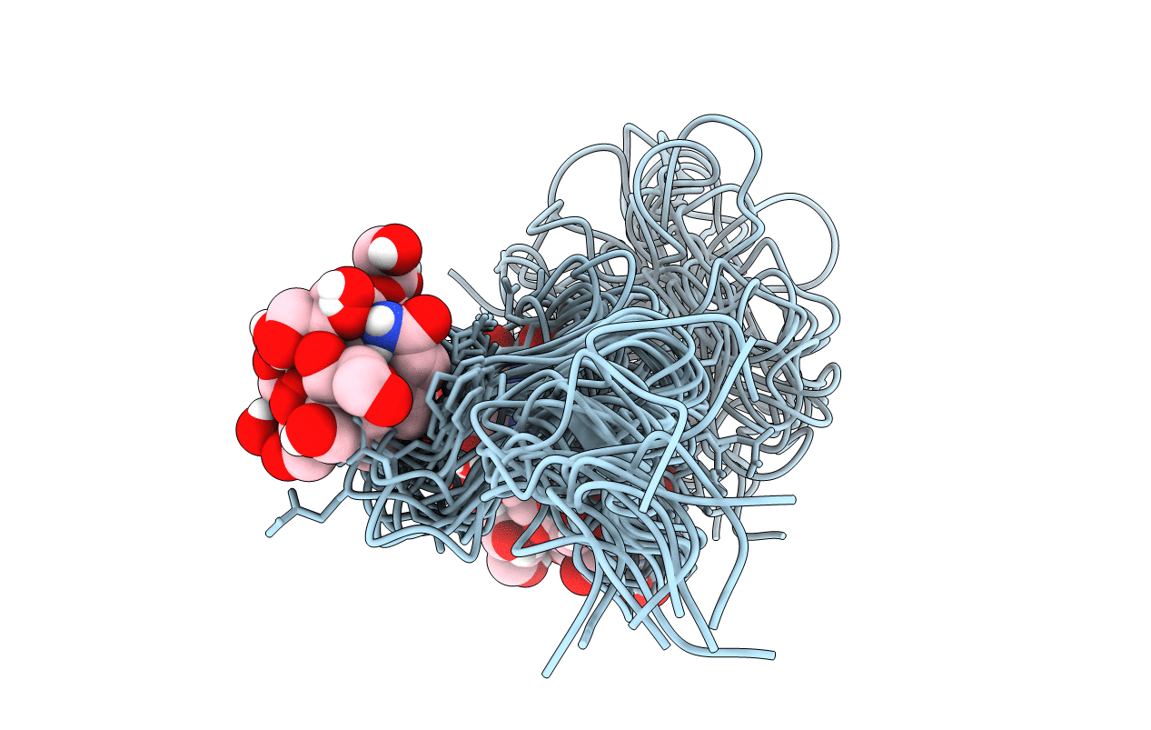
1DQB image
Deposition Date 2000-01-03
Release Date 2000-03-06
Last Version Date 2024-10-16
Entry Detail
PDB ID:
1DQB
Title:
NMR STRUCTURE OF THROMBOMODULIN EGF(4-5)
Biological Source:
Source Organism:
Homo sapiens (Taxon ID: 9606)
Host Organism:
Method Details:
Experimental Method:
Conformers Calculated:
50
Conformers Submitted:
12
Selection Criteria:
structures with the least restraint violations
Macromolecular Entities
Structures with similar UniProt ID
Protein Blast
Polymer Type:polypeptide(L)
Molecule:THROMBOMODULIN
Gene (Uniprot):THBD
Chain IDs:A
Chain Length:83
Number of Molecules:1
Biological Source:Homo sapiens
Modified Residue
Compound ID Chain ID Parent Comp ID Details 2D Image
ASN A ASN GLYCOSYLATION SITE
Ligand Molecules
Primary Citation
Solution structure of the smallest cofactor-active fragment of thrombomodulin.
Nat.Struct.Biol. 7 200 204 (2000)
PMID: 10700277 DOI: 10.1038/73302

Abstact

A glycosylated fragment of thrombomodulin containing two epidermal growth factor-like domains (TMEGF45) was analyzed by NMR. The 4th-domains structure of this two-domain fragment is similar to that of the individual domain previously determined. The 5th-domain, which has uncrossed disulfide bonds, is not as well determined in the two-domain fragment than the individual domain previously solved. The flexibility of the 5th-domain is consistent with low heteronuclear NOEs. In the individual 5th-domain, Met 388 was disordered, and key thrombin binding residues formed a hydrophobic core. By contrast, in TMEGF45, Met 388 is in the 5th-domain core, positioned by Phe 376 from the 4th-domain. As a result, key thrombin binding residues that were in the core of the individual domain are expelled. Upon thrombin binding, chemical shifts of two residues in the 4th-domain, the three interdomain linker residues, and nearly all of the 5th-domain are perturbed. Thus, TMEGF45 binds thrombin by an induced fit mechanism involving a flexible 5th-domain.

Legend

Protein

Chemical

Disease

Primary Citation of related structures