1DOW image
Deposition Date 1999-12-21
Release Date 2000-07-12
Last Version Date 2024-11-13
Entry Detail
PDB ID:
1DOW
Keywords:
Title:
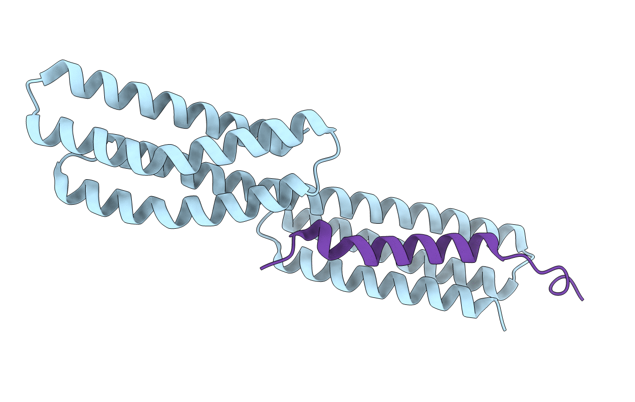
CRYSTAL STRUCTURE OF A CHIMERA OF BETA-CATENIN AND ALPHA-CATENIN
Biological Source:
Source Organism:
Mus musculus (Taxon ID: 10090)
Host Organism:
Method Details:
Experimental Method:
Resolution:
1.80 Å
R-Value Free:
0.23
R-Value Work:
0.2
R-Value Observed:
0.2
Space Group:
P 21 21 21
Macromolecular Entities
Polymer Type:polypeptide(L)
Molecule:ALPHA-CATENIN
Gene (Uniprot):Ctnna1
Chain IDs:A
Chain Length:205
Number of Molecules:1
Biological Source:Mus musculus
Polymer Type:polypeptide(L)
Molecule:BETA-CATENIN
Gene (Uniprot):Ctnnb1
Chain IDs:B
Chain Length:32
Number of Molecules:1
Biological Source:Mus musculus
Modified Residue
Compound ID Chain ID Parent Comp ID Details 2D Image
MSE A MET SELENOMETHIONINE
Primary Citation
Structure of the dimerization and beta-catenin-binding region of alpha-catenin.
Mol.Cell 5 533 543 (2000)
PMID: 10882138 DOI: 10.1016/S1097-2765(00)80447-5

Abstact

In adherens junctions, alpha-catenin links the cadherin-beta-catenin complex to the actin-based cytoskeleton. alpha-catenin is a homodimer in solution, but forms a 1:1 heterodimer with beta-catenin. The crystal structure of the alpha-catenin dimerization domain, residues 82-279, shows that alpha-catenin dimerizes through formation of a four-helix bundle in which two antiparallel helices are contributed by each protomer. A slightly larger fragment, comprising residues 57-264, binds to beta-catenin. A chimera consisting of the alpha-catenin-binding region of beta-catenin linked to the amino terminus of alpha-catenin 57-264 behaves as a monomer in solution, as expected, since beta-catenin binding disrupts the alpha-catenin dimer. The crystal structure of this chimera reveals the interaction between alpha- and beta-catenin, and provides a basis for understanding adherens junction assembly.

Legend

Protein

Chemical

Disease

Primary Citation of related structures