Deposition Date
1993-01-12
Release Date
1994-01-31
Last Version Date
2024-10-23
Entry Detail
PDB ID:
1DOG
Keywords:
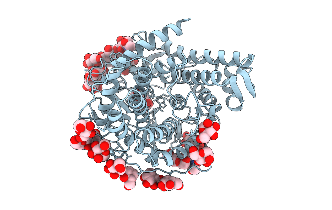
Title:
REFINED STRUCTURE FOR THE COMPLEX OF 1-DEOXYNOJIRIMYCIN WITH GLUCOAMYLASE FROM (ASPERGILLUS AWAMORI) VAR. X100 TO 2.4 ANGSTROMS RESOLUTION
Biological Source:
Source Organism(s):
Aspergillus awamori (Taxon ID: 105351)
Method Details:
Experimental Method:
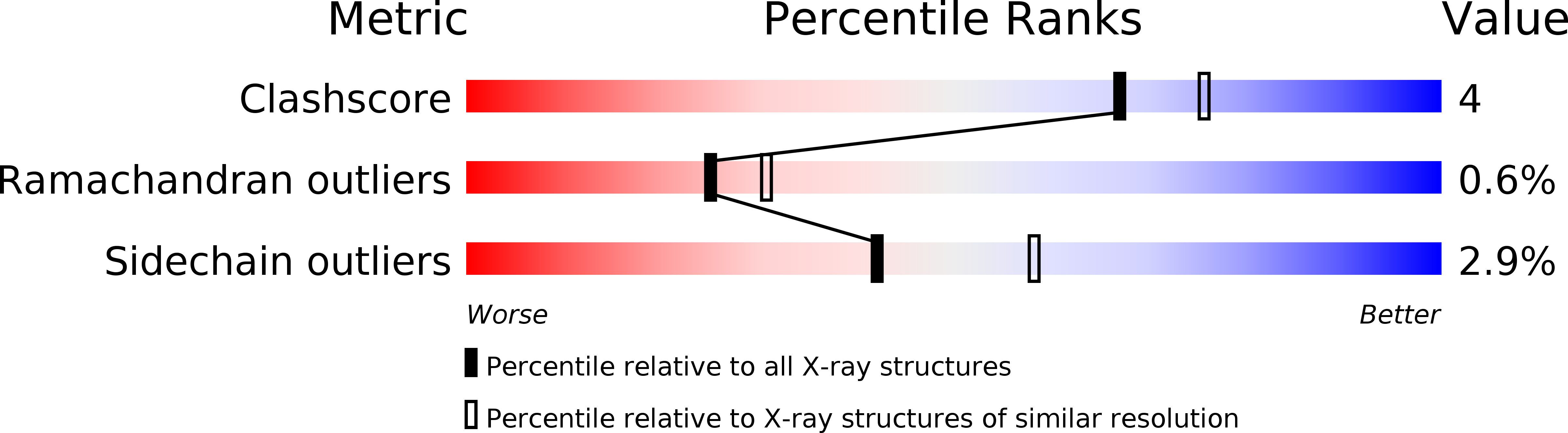
Resolution:
2.30 Å
R-Value Observed:
0.11
Space Group:
P 21 21 21