
Deposition Date
1999-12-18
Release Date
2000-01-05
Last Version Date
2024-11-06
Entry Detail
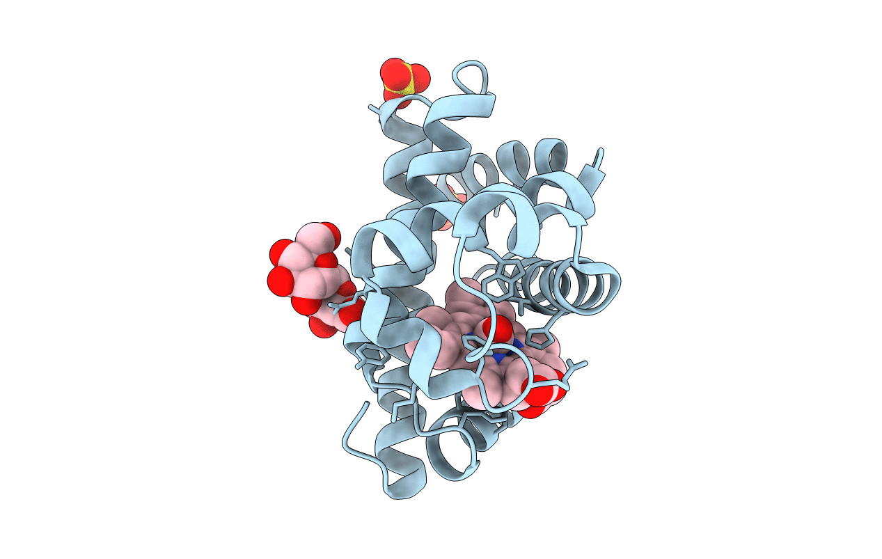
Biological Source:
Source Organism(s):
Physeter catodon (Taxon ID: 9755)
Expression System(s):
Method Details:
Experimental Method:
Resolution:
1.50 Å
R-Value Free:
0.21
R-Value Work:
0.18
R-Value Observed:
0.19
Space Group:
P 6
