1DNL image
Deposition Date 1999-12-16
Release Date 2000-01-05
Last Version Date 2024-10-09
Entry Detail
PDB ID:
1DNL
Keywords:
Title:
X-RAY STRUCTURE OF ESCHERICHIA COLI PYRIDOXINE 5'-PHOSPHATE OXIDASE COMPLEXED WITH FMN AT 1.8 ANGSTROM RESOLUTION
Biological Source:
Source Organism:
Host Organism:
Method Details:
Experimental Method:
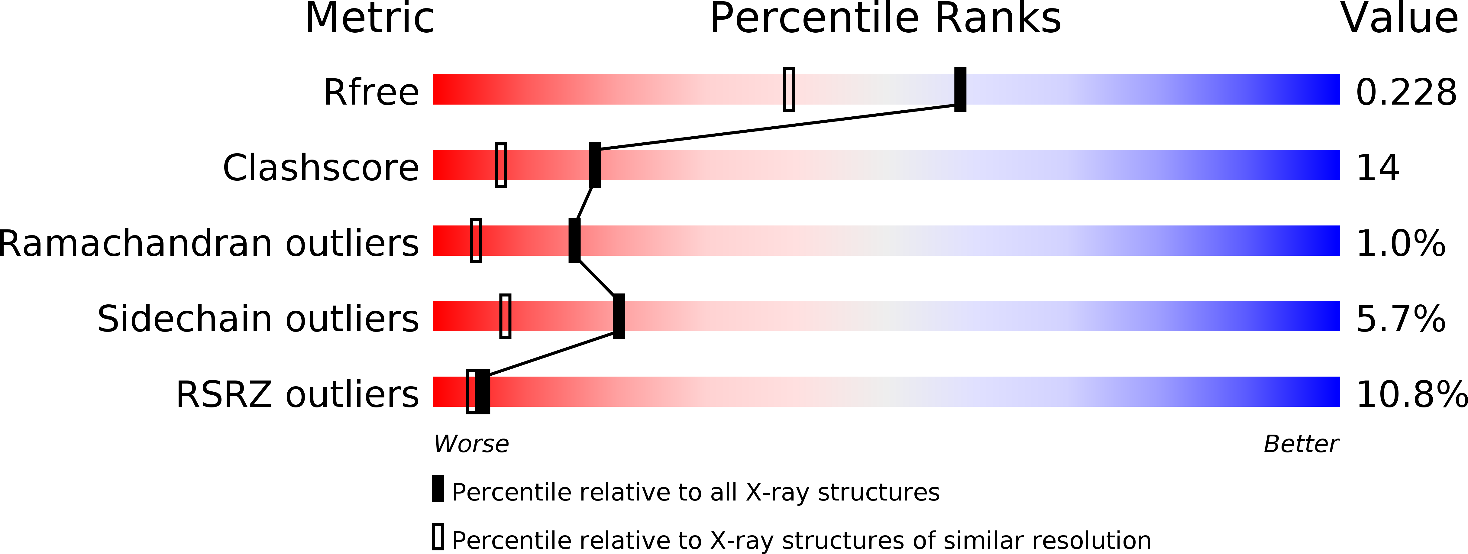
Resolution:
1.80 Å
R-Value Free:
0.23
R-Value Work:
0.21
R-Value Observed:
0.21
Space Group:
P 31 2 1
Macromolecular Entities
Structures with similar UniProt ID
Protein Blast
Polymer Type:polypeptide(L)
Molecule:PYRIDOXINE 5'-PHOSPHATE OXIDASE
Chain IDs:A
Chain Length:199
Number of Molecules:1
Biological Source:Escherichia coli K12
Modified Residue
Compound ID Chain ID Parent Comp ID Details 2D Image
MSE A MET SELENOMETHIONINE
Primary Citation
X-ray structure of Escherichia coli pyridoxine 5'-phosphate oxidase complexed with FMN at 1.8 A resolution.
Structure Fold.Des. 8 751 762 (2000)
PMID: 10903950 DOI: 10.1016/S0969-2126(00)00162-3

Abstact

BACKGROUND: Escherichia coli pyridoxine 5'-phosphate oxidase (PNPOx) catalyzes the terminal step in the biosynthesis of pyridoxal 5'-phosphate (PLP), a cofactor used by many enzymes involved in amino acid metabolism. The enzyme oxidizes either the 4'-hydroxyl group of pyridoxine 5'-phosphate (PNP) or the 4'-primary amine of pyridoxamine 5'-phosphate (PMP) to an aldehyde. PNPOx is a homodimeric enzyme with one flavin mononucleotide (FMN) molecule non-covalently bound to each subunit. A high degree of sequence homology among the 15 known members of the PNPOx family suggests that all members of this group have similar three-dimensional folds. RESULTS: The crystal structure of PNPOx from E. coli has been determined to 1.8 A resolution. The monomeric subunit folds into an eight-stranded beta sheet surrounded by five alpha-helical structures. Two monomers related by a twofold axis interact extensively along one-half of each monomer to form the dimer. There are two clefts at the dimer interface that are symmetry-related and extend from the top to the bottom of the dimer. An FMN cofactor that makes interactions with both subunits is located in each of these two clefts. CONCLUSIONS: The structure is quite similar to the recently deposited 2.7 A structure of Saccharomyces cerevisiae PNPOx and also, remarkably, shares a common structural fold with the FMN-binding protein from Desulfovibrio vulgaris and a domain of chymotrypsin. This high-resolution E. coli PNPOx structure permits predictions to be made about residues involved in substrate binding and catalysis. These predictions provide testable hypotheses, which can be answered by making site-directed mutants.

Legend

Protein

Chemical

Disease

Primary Citation of related structures