1DKF image
Deposition Date 1999-12-07
Release Date 2000-04-19
Last Version Date 2024-02-07
Entry Detail
PDB ID:
1DKF
Title:
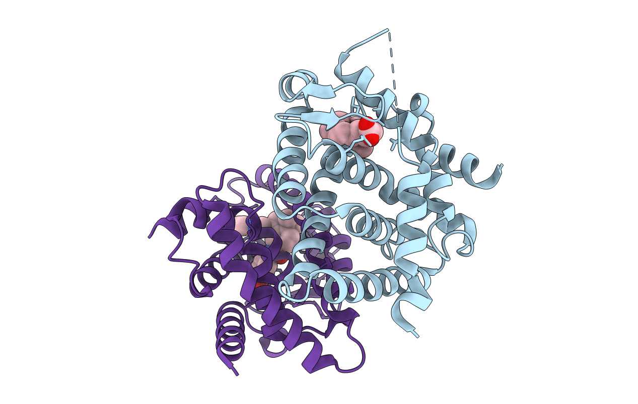
CRYSTAL STRUCTURE OF A HETERODIMERIC COMPLEX OF RAR AND RXR LIGAND-BINDING DOMAINS
Biological Source:
Source Organism:
Mus musculus (Taxon ID: 10090)
Homo sapiens (Taxon ID: 9606)
Host Organism:
Method Details:
Experimental Method:
Resolution:
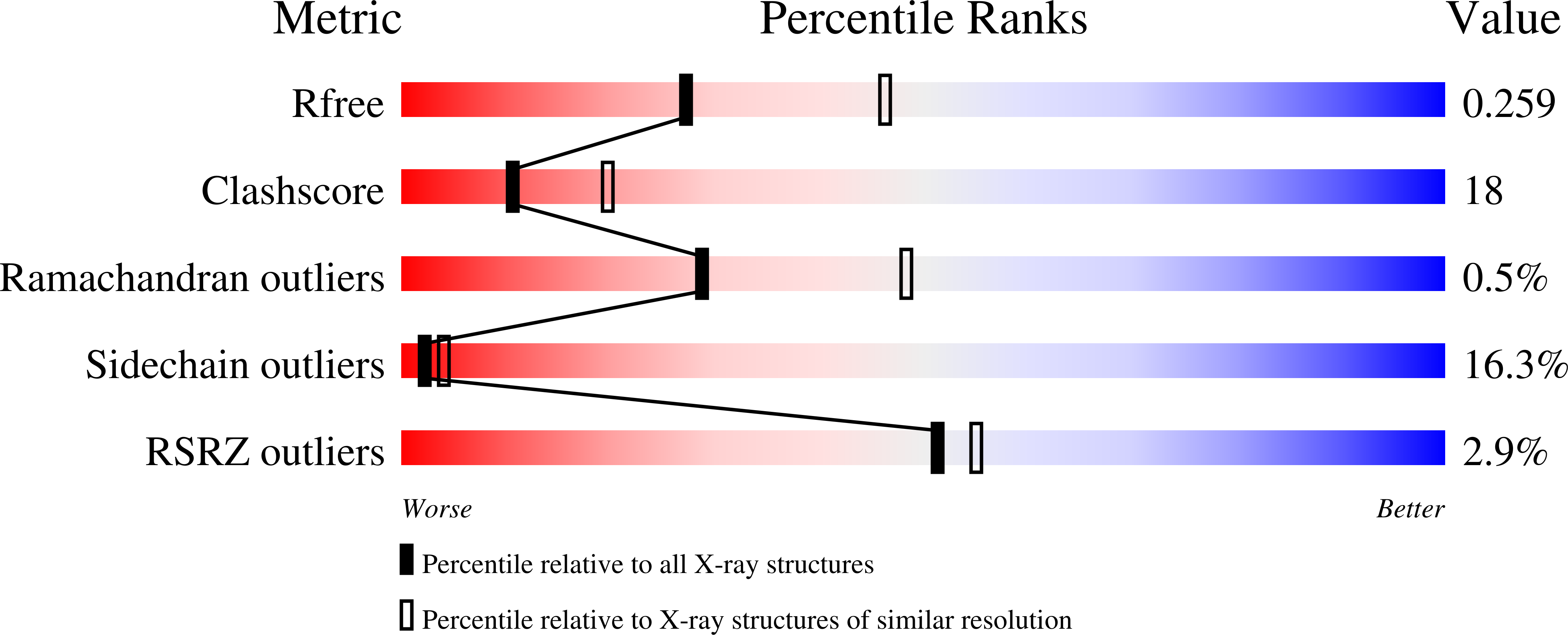
2.50 Å
R-Value Free:
0.26
R-Value Work:
0.20
R-Value Observed:
0.20
Space Group:
P 61 2 2
Macromolecular Entities
Structures with similar UniProt ID
Protein Blast
Polymer Type:polypeptide(L)
Molecule:PROTEIN (RETINOID X RECEPTOR-ALPHA)
Gene (Uniprot):Rxra
Mutagens:YES
Chain IDs:A
Chain Length:233
Number of Molecules:1
Biological Source:Mus musculus
Structures with similar UniProt ID
Protein Blast
Polymer Type:polypeptide(L)
Molecule:PROTEIN (RETINOIC ACID RECEPTOR-ALPHA)
Gene (Uniprot):RARA
Chain IDs:B
Chain Length:235
Number of Molecules:1
Biological Source:Homo sapiens
Primary Citation
Crystal structure of a heterodimeric complex of RAR and RXR ligand-binding domains.
Mol.Cell 5 289 298 (2000)
PMID: 10882070 DOI: 10.1016/S1097-2765(00)80424-4

Abstact

The crystal structure of a heterodimer between the ligand-binding domains (LBDs) of the human RARalpha bound to a selective antagonist and the constitutively active mouse RXRalphaF318A mutant shows that, pushed by a bulky extension of the ligand, RARalpha helix H12 adopts an antagonist position. The unexpected presence of a fatty acid in the ligand-binding pocket of RXRalpha(F318A is likely to account for its apparent "constitutivity." Specific conformational changes suggest the structural basis of pure and partial antagonism. The RAR-RXR heterodimer interface is similar to that observed in most nuclear receptor (NR) homodimers. A correlative analysis of 3D structures and sequences provides a novel view on dimerization among members of the nuclear receptor superfamily.

Legend

Protein

Chemical

Disease

Primary Citation of related structures