Deposition Date
1996-09-25
Release Date
1997-07-07
Last Version Date
2024-02-07
Entry Detail
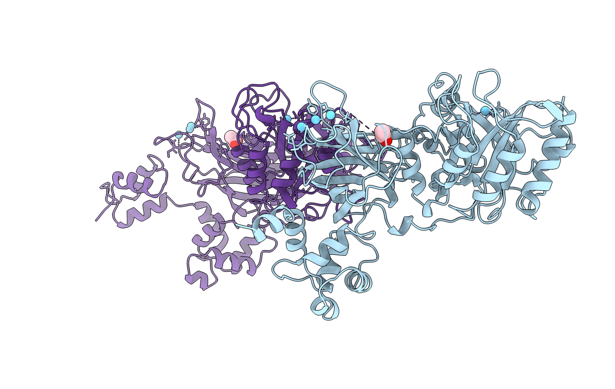
PDB ID:
1DJG
Keywords:
Title:
PHOSPHOINOSITIDE-SPECIFIC PHOSPHOLIPASE C-DELTA1 FROM RAT COMPLEXED WITH LANTHANUM
Biological Source:
Source Organism:
Rattus norvegicus (Taxon ID: 10116)
Host Organism:
Method Details:
Experimental Method:
Resolution:
2.60 Å
R-Value Free:
0.27
R-Value Work:
0.21
Space Group:
F 41 3 2