Deposition Date
1992-03-30
Release Date
1993-07-15
Last Version Date
2024-02-07
Entry Detail
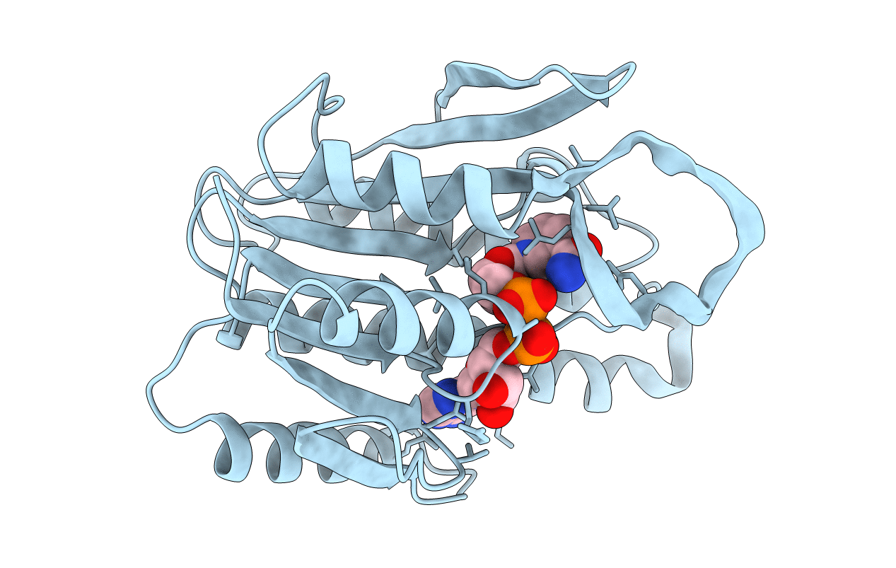
PDB ID:
1DHR
Keywords:
Title:
CRYSTAL STRUCTURE OF RAT LIVER DIHYDROPTERIDINE REDUCTASE
Biological Source:
Source Organism(s):
Rattus norvegicus (Taxon ID: 10116)
Method Details: