Deposition Date
1999-11-17
Release Date
2000-03-09
Last Version Date
2024-02-07
Entry Detail
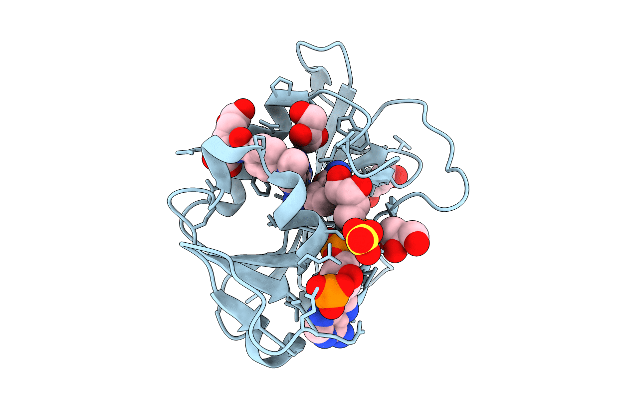
PDB ID:
1DF7
Keywords:
Title:
DIHYDROFOLATE REDUCTASE OF MYCOBACTERIUM TUBERCULOSIS COMPLEXED WITH NADPH AND METHOTREXATE
Biological Source:
Source Organism(s):
Mycobacterium tuberculosis (Taxon ID: 1773)
Method Details:
Experimental Method:
Resolution:
1.70 Å
R-Value Free:
0.24
R-Value Work:
0.18
R-Value Observed:
0.18
Space Group:
P 41