Deposition Date
1999-11-15
Release Date
2000-02-02
Last Version Date
2023-08-09
Entry Detail
PDB ID:
1DEQ
Keywords:
Title:
THE CRYSTAL STRUCTURE OF MODIFIED BOVINE FIBRINOGEN (AT ~4 ANGSTROM RESOLUTION)
Biological Source:
Source Organism:
Bos taurus (Taxon ID: 9913)
Method Details:
Experimental Method:
Resolution:
3.50 Å
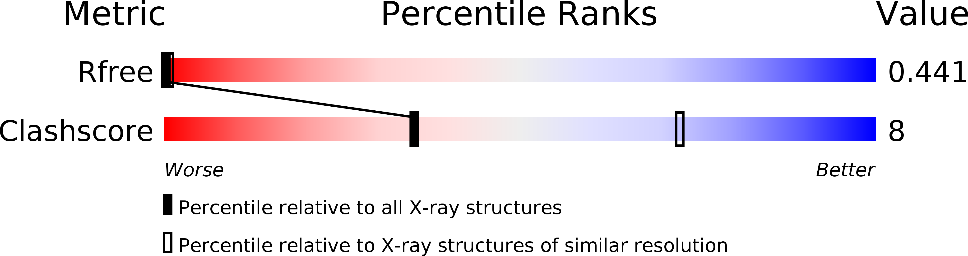
R-Value Free:
0.37
R-Value Work:
0.25
R-Value Observed:
0.26
Space Group:
P 1 21 1