Deposition Date
1994-03-01
Release Date
1994-07-31
Last Version Date
2024-11-20
Entry Detail
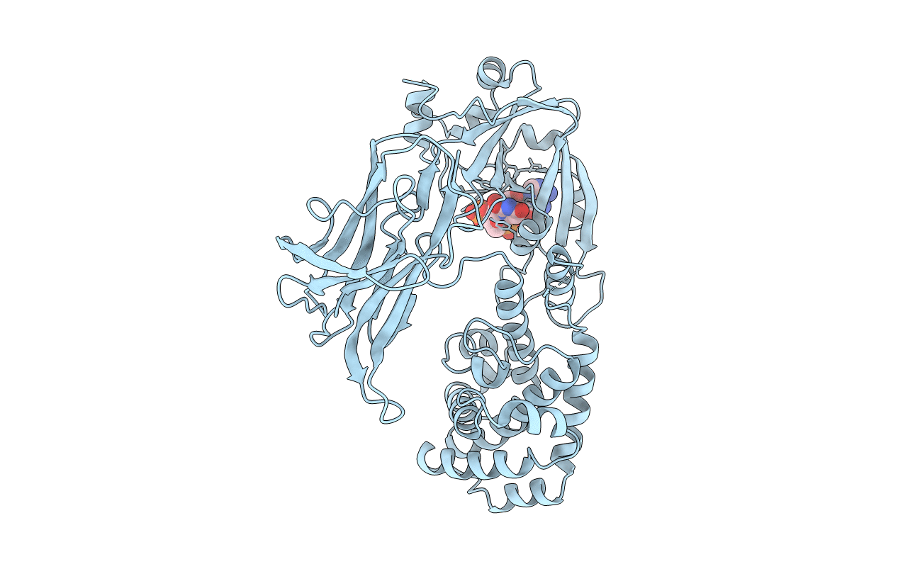
PDB ID:
1DDT
Keywords:
Title:
THE REFINED STRUCTURE OF DIMERIC DIPHTHERIA TOXIN AT 2.0 ANGSTROMS RESOLUTION
Biological Source:
Source Organism(s):
Corynephage beta (Taxon ID: 10703)
Method Details:
Experimental Method:
Resolution:
2.00 Å
R-Value Work:
0.19
R-Value Observed:
0.19
Space Group:
C 1 2 1