1DDB image
Deposition Date 1999-02-19
Release Date 1999-08-30
Last Version Date 2023-12-27
Entry Detail
PDB ID:
1DDB
Keywords:
Title:
STRUCTURE OF MOUSE BID, NMR, 20 STRUCTURES
Biological Source:
Source Organism:
Mus musculus (Taxon ID: 10090)
Method Details:
Experimental Method:
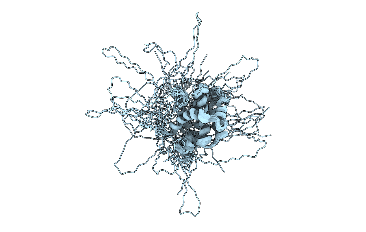
Conformers Calculated:
1000
Conformers Submitted:
20
Selection Criteria:
LEAST RESTRAINT VIOLATION
Macromolecular Entities
Structures with similar UniProt ID
Protein Blast
Polymer Type:polypeptide(L)
Molecule:PROTEIN (BID)
Gene (Uniprot):Bid
Chain IDs:A
Chain Length:195
Number of Molecules:1
Biological Source:Mus musculus
Ligand Molecules
Primary Citation
Solution structure of the proapoptotic molecule BID: a structural basis for apoptotic agonists and antagonists.
Cell(Cambridge,Mass.) 96 625 634 (1999)
PMID: 10089878 DOI: 10.1016/S0092-8674(00)80573-5

Abstact

Members of the BCL2 family of proteins are key regulators of programmed cell death, acting either as apoptotic agonists or antagonists. Here we describe the solution structure of BID, presenting the structure of a proapoptotic BCL2 family member. An analysis of sequence/structure of BCL2 family members allows us to define a structural superfamily, which has implications for general mechanisms for regulating proapoptotic activity. It appears two criteria must be met for proapoptotic function within the BCL2 family: targeting of molecules to intracellular membranes, and exposure of the BH3 death domain. BID's activity is regulated by a Caspase 8-mediated cleavage event, exposing the BH3 domain and significantly changing the surface charge and hydrophobicity, resulting in a change of cellular localization.

Legend

Protein

Chemical

Disease

Primary Citation of related structures