
Deposition Date
1999-11-04
Release Date
2000-03-24
Last Version Date
2024-10-16
Entry Detail
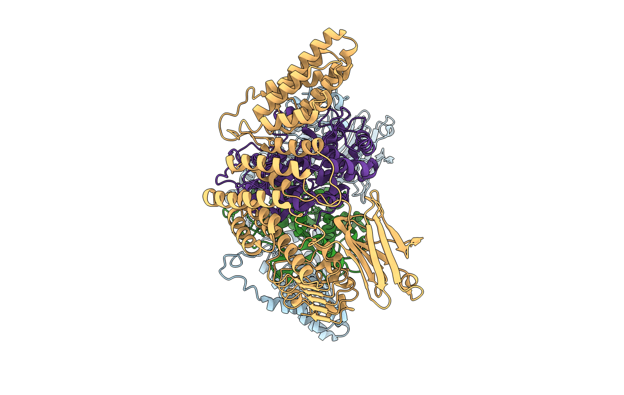
PDB ID:
1DCE
Keywords:
Title:
CRYSTAL STRUCTURE OF RAB GERANYLGERANYLTRANSFERASE FROM RAT BRAIN
Biological Source:
Source Organism(s):
Rattus norvegicus (Taxon ID: 10116)
Method Details:
Experimental Method:
Resolution:
2.00 Å
R-Value Free:
0.26
R-Value Work:
0.21
Space Group:
P 1
