Deposition Date
1999-11-04
Release Date
2001-02-21
Last Version Date
2024-02-07
Entry Detail
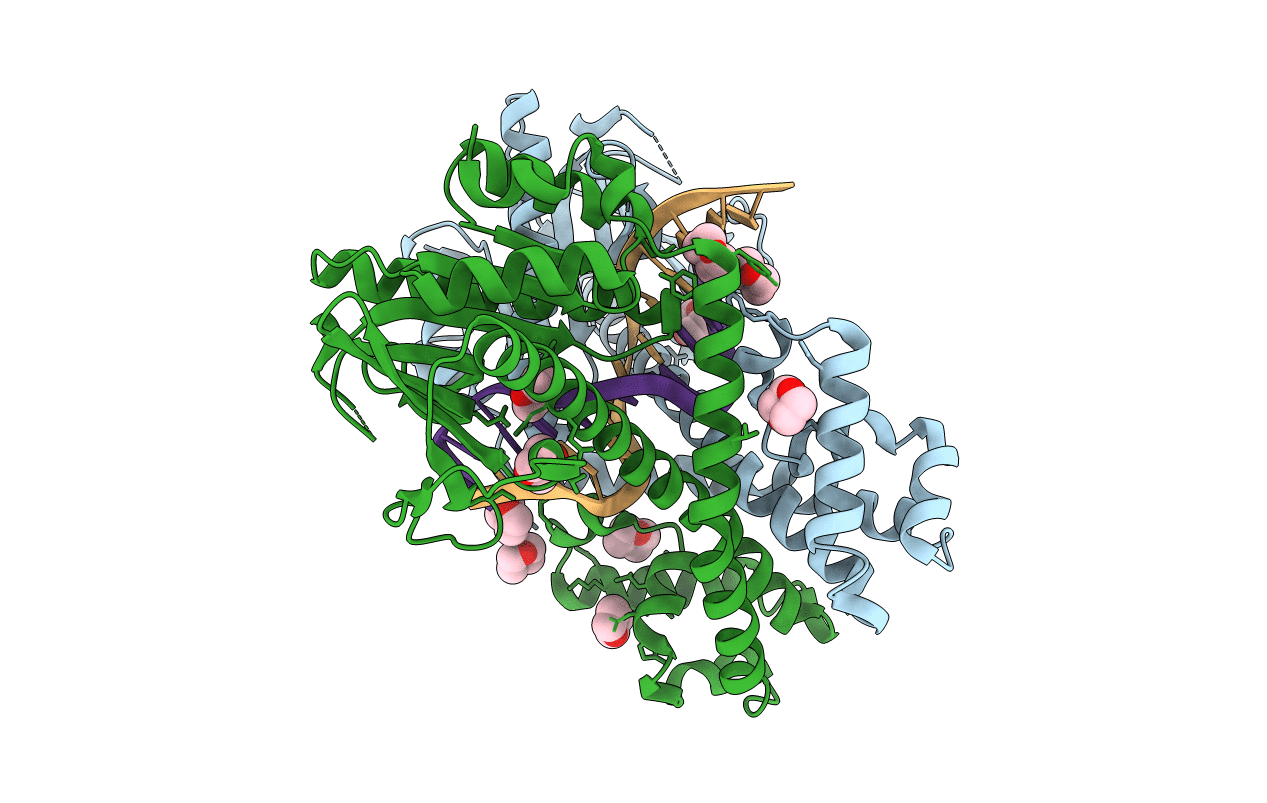
PDB ID:
1DC1
Keywords:
Title:
RESTRICTION ENZYME BSOBI/DNA COMPLEX STRUCTURE: ENCIRCLEMENT OF THE DNA AND HISTIDINE-CATALYZED HYDROLYSIS WITHIN A CANONICAL RESTRICTION ENZYME FOLD
Biological Source:
Source Organism(s):
Geobacillus stearothermophilus (Taxon ID: 1422)
Expression System(s):
Method Details:
Experimental Method:
Resolution:
1.70 Å
R-Value Free:
0.25
R-Value Work:
0.19
R-Value Observed:
0.19
Space Group:
P 21 21 21