Deposition Date
1999-11-03
Release Date
1999-11-26
Last Version Date
2024-02-07
Entry Detail
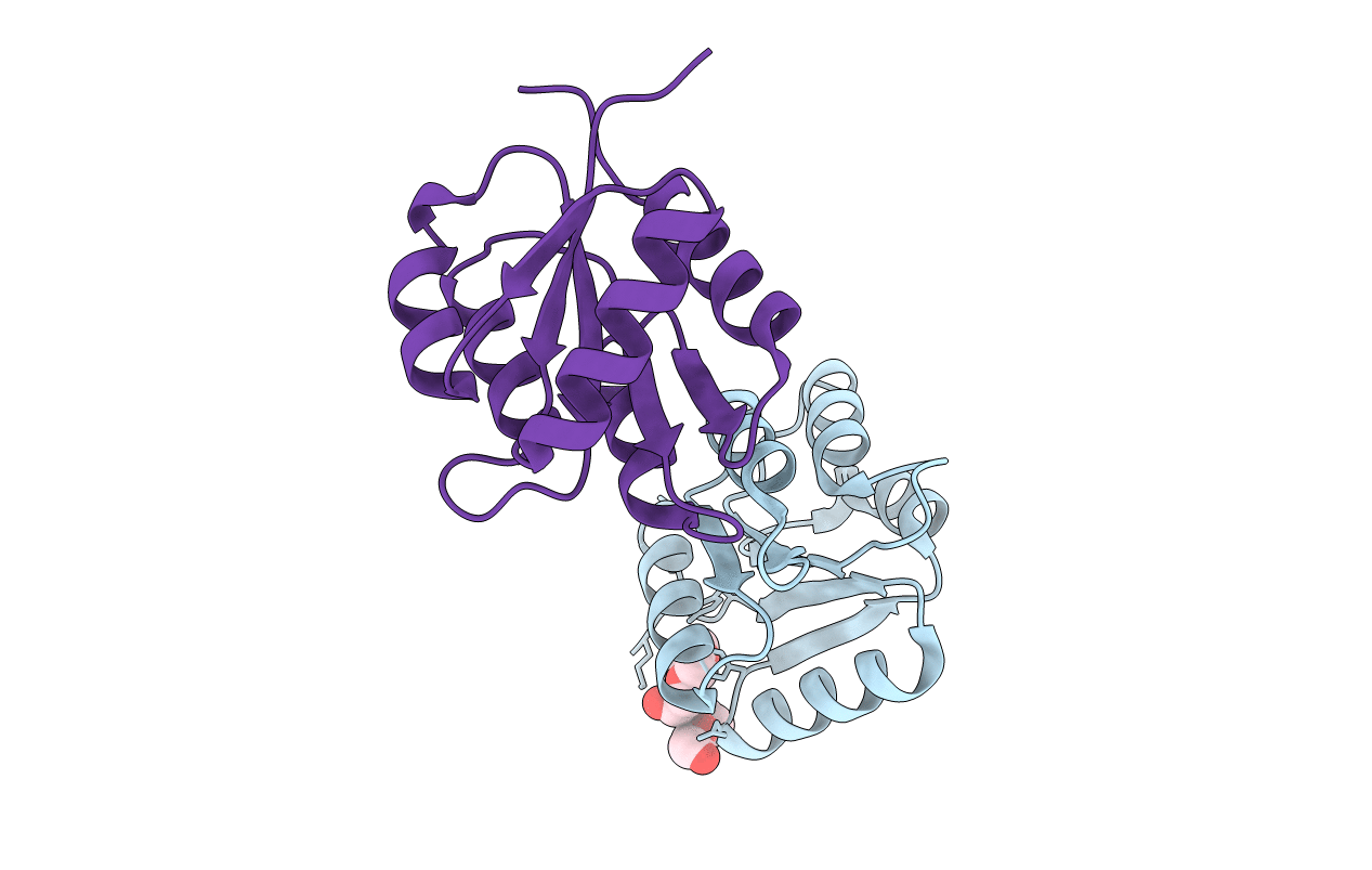
Biological Source:
Source Organism(s):
Sinorhizobium meliloti (Taxon ID: 382)
Expression System(s):
Method Details:
Experimental Method:
Resolution:
1.60 Å
R-Value Free:
0.22
R-Value Work:
0.18
R-Value Observed:
0.18
Space Group:
P 1