
Deposition Date
1998-01-21
Release Date
1998-05-27
Last Version Date
2024-05-22
Entry Detail
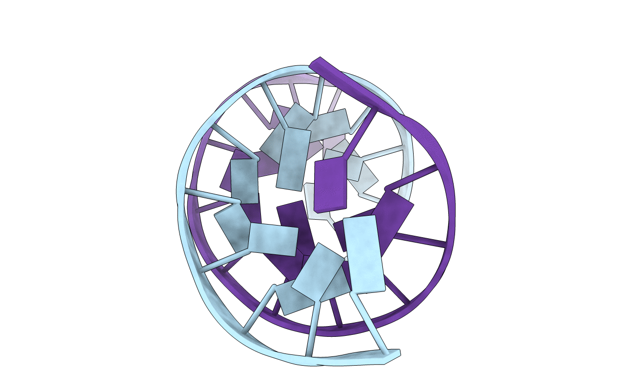
PDB ID:
1DAU
Keywords:
Title:
Analog of dickerson-drew DNA dodecamer with 6'-alpha-methyl carbocyclic thymidines, NMR, minimized average structure
Method Details:
Experimental Method:
Conformers Calculated:
9
Conformers Submitted:
1
Selection Criteria:
LOWEST ENERGY, BEST AGREEMENT WITH NOE VOLUMES
