1D9X image
Deposition Date 1999-10-30
Release Date 2000-05-03
Last Version Date 2024-02-07
Entry Detail
PDB ID:
1D9X
Keywords:
Title:
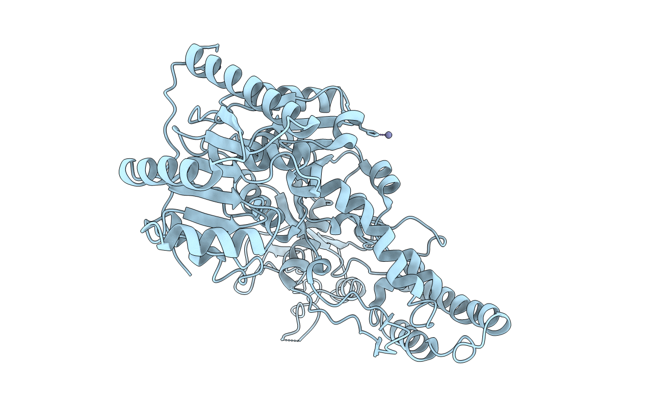
CRYSTAL STRUCTURE OF THE DNA REPAIR PROTEIN UVRB
Biological Source:
Source Organism:
Host Organism:
Method Details:
Experimental Method:
Resolution:
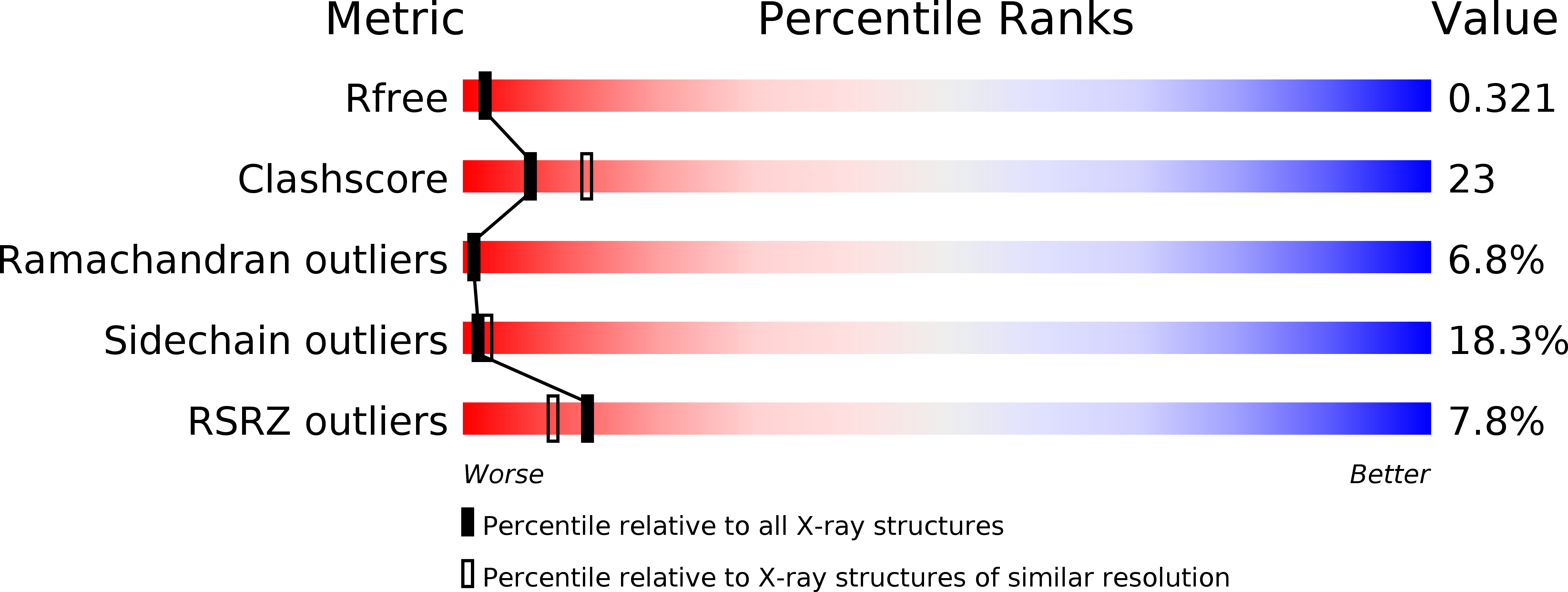
2.60 Å
R-Value Free:
0.32
R-Value Work:
0.25
Space Group:
P 31 2 1
Macromolecular Entities
Polymer Type:polypeptide(L)
Molecule:EXCINUCLEASE UVRABC COMPONENT UVRB
Gene (Uniprot):uvrB
Chain IDs:A
Chain Length:658
Number of Molecules:1
Biological Source:Bacillus caldotenax
Ligand Molecules
Primary Citation
Crystal structure of UvrB, a DNA helicase adapted for nucleotide excision repair.
EMBO J. 18 6899 6907 (1999)
PMID: 10601012 DOI: 10.1093/emboj/18.24.6899

Abstact

Nucleotide excision repair (NER) is a highly conserved DNA repair mechanism. NER systems recognize the damaged DNA strand, cleave it on both sides of the lesion, remove and newly synthesize the fragment. UvrB is a central component of the bacterial NER system participating in damage recognition, strand excision and repair synthesis. We have solved the crystal structure of UvrB in the apo and the ATP-bound forms. UvrB contains two domains related in structure to helicases, and two additional domains unique to repair proteins. The structure contains all elements of an intact helicase, and is evidence that UvrB utilizes ATP hydrolysis to move along the DNA to probe for damage. The location of conserved residues and structural comparisons allow us to predict the path of the DNA and suggest that the tight pre-incision complex of UvrB and the damaged DNA is formed by insertion of a flexible beta-hairpin between the two DNA strands.

Legend

Protein

Chemical

Disease

Primary Citation of related structures