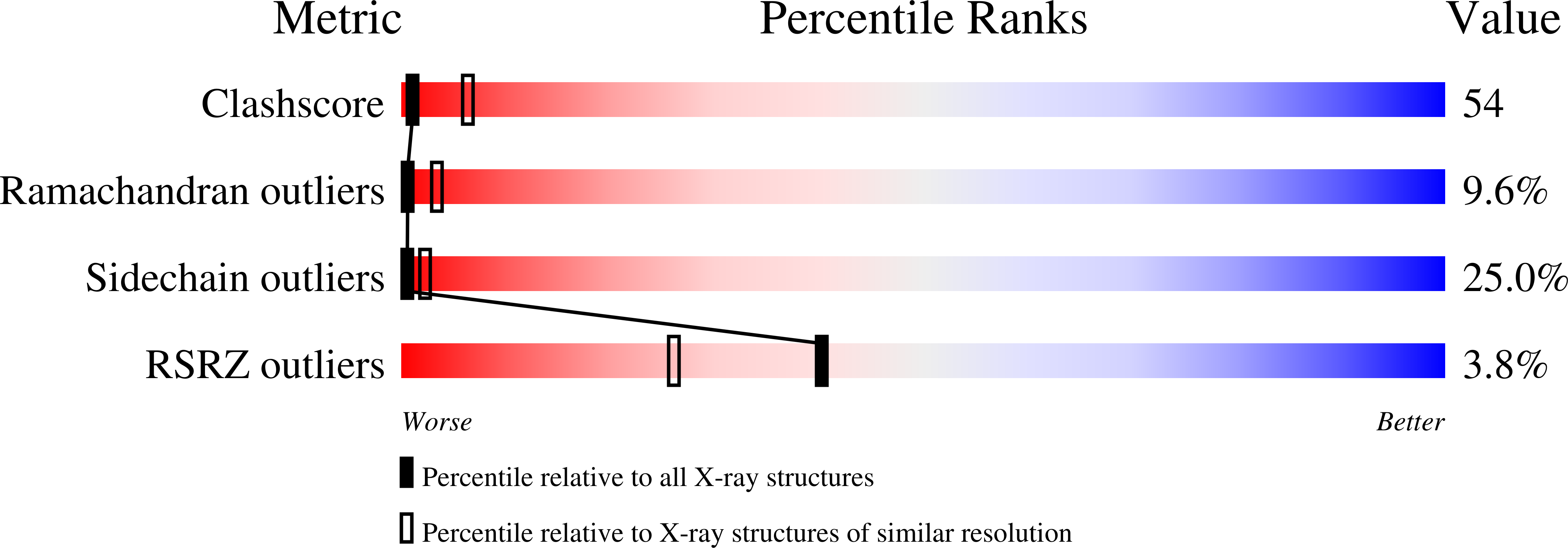
1D9K image
Deposition Date 1999-10-28
Release Date 1999-12-15
Last Version Date 2024-11-20
Entry Detail
PDB ID:
1D9K
Keywords:
Title:
CRYSTAL STRUCTURE OF COMPLEX BETWEEN D10 TCR AND PMHC I-AK/CA
Biological Source:
Source Organism:
Mus musculus (Taxon ID: 10090)
Host Organism:
Method Details:
Experimental Method:
Resolution:
3.20 Å
R-Value Free:
0.29
R-Value Work:
0.24
R-Value Observed:
0.07
Space Group:
P 21 21 2
Macromolecular Entities
Polymer Type:polypeptide(L)
Molecule:T-CELL RECEPTOR D10 (ALPHA CHAIN)
Mutations:C115S
Chain IDs:A, F (auth: E)
Chain Length:110
Number of Molecules:2
Biological Source:Mus musculus
Polymer Type:polypeptide(L)
Molecule:T-CELL RECEPTOR D10 (BETA CHAIN)
Chain IDs:B, G (auth: F)
Chain Length:112
Number of Molecules:2
Biological Source:Mus musculus
Polymer Type:polypeptide(L)
Molecule:MHC I-AK A CHAIN (ALPHA CHAIN)
Gene (Uniprot):H2-Aa
Chain IDs:C, H (auth: G)
Chain Length:183
Number of Molecules:2
Biological Source:Mus musculus
Polymer Type:polypeptide(L)
Molecule:MHC I-AK B CHAIN (BETA CHAIN)
Gene (Uniprot):H2-Ab1
Chain IDs:D, I (auth: H)
Chain Length:188
Number of Molecules:2
Biological Source:Mus musculus
Polymer Type:polypeptide(L)
Molecule:CONALBUMIN PEPTIDE
Chain IDs:E (auth: P), J (auth: Q)
Chain Length:16
Number of Molecules:2
Biological Source:Mus musculus
Modified Residue
Compound ID Chain ID Parent Comp ID Details 2D Image
ASN C ASN GLYCOSYLATION SITE
Ligand Molecules
Primary Citation
The crystal structure of a T cell receptor in complex with peptide and MHC class II.
Science 286 1913 1921 (1999)
PMID: 10583947 DOI: 10.1126/science.286.5446.1913

Abstact

The crystal structure of a complex involving the D10 T cell receptor (TCR), 16-residue foreign peptide antigen, and the I-Ak self major histocompatibility complex (MHC) class II molecule is reported at 3.2 angstrom resolution. The D10 TCR is oriented in an orthogonal mode relative to its peptide-MHC (pMHC) ligand, necessitated by the amino-terminal extension of peptide residues projecting from the MHC class II antigen-binding groove as part of a mini beta sheet. Consequently, the disposition of D10 complementarity-determining region loops is altered relative to that of most pMHCI-specific TCRs; the latter TCRs assume a diagonal orientation, although with substantial variability. Peptide recognition, which involves P-1 to P8 residues, is dominated by the Valpha domain, which also binds to the class II MHC beta1 helix. That docking is limited to one segment of MHC-bound peptide offers an explanation for epitope recognition and altered peptide ligand effects, suggests a structural basis for alloreactivity, and illustrates how bacterial superantigens can span the TCR-pMHCII surface.

Legend

Protein

Chemical

Disease

Primary Citation of related structures