1D81 image
Deposition Date 1992-07-07
Release Date 1992-10-15
Last Version Date 2024-02-07
Entry Detail
PDB ID:
1D81
Keywords:
Title:
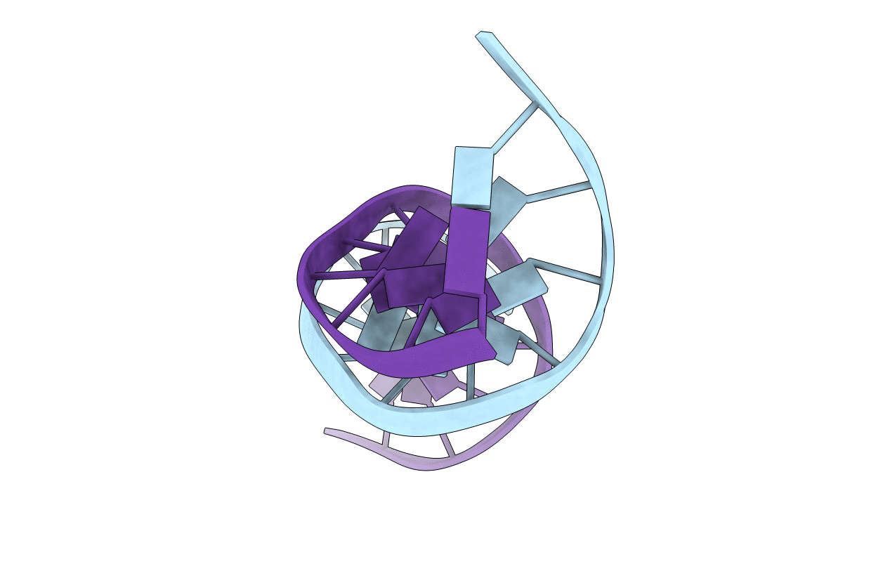
THE CONFORMATIONAL VARIABILITY OF AN ADENOSINE. INOSINE BASE-PAIR IN A SYNTHETIC DNA DODECAMER
Biological Source:
Source Organism:
Method Details:
Experimental Method:
Resolution:
2.50 Å
R-Value Observed:
0.15
Space Group:
P 21 21 21
Macromolecular Entities
Polymer Type:polydeoxyribonucleotide
Molecule:DNA (5'-D(*CP*GP*CP*AP*AP*AP*TP*TP*IP*GP*CP*G)-3')
Chain IDs:A, B
Chain Length:12
Number of Molecules:2
Biological Source:
Primary Citation
The conformational variability of an adenosine.inosine base-pair in a synthetic DNA dodecamer.
Nucleic Acids Res. 20 4753 4759 (1992)
PMID: 1408788 DOI: 10.1093/nar/20.18.4753

Abstact

A crystal structure analysis of the synthetic deoxydodecamer d(CGCAAATTIGCG) which contains two adenosine.inosine (A.I) mispairs has revealed that, in this sequence, the A.I base-pairs adopt a A(anti).I(syn) configuration. The refinement converged at R = 0.158 for 2004 reflections with F greater than or equal to 2 sigma(F) in the range 7.0-2.5A for a model consisting of the DNA duplex and 71 water molecules. A notable feature of the structure is the presence of an almost complete spine of hydration spanning the minor groove of the whole of the (AAATTI)2 core region of the duplex. pH-dependent ultraviolet melting studies have suggested that the base-pair observed in the crystal structure is, in fact, a protonated AH+ (anti).I(syn) species and that the A.I base-pairs in the sequence studied display the same conformational variability as A.G mispairs in the sequence d(CGCAAATTGGCG). The AH+(anti).I(syn) base-pair predominates below pH 6.5 and an A(anti).I(anti) mispair is the major species present between pH 6.5 and 8.0. The protonated base-pairs are held together by two hydrogen bonds one between N6(A) and O6(I) and the other between N1(A) and N7(I). This second hydrogen bond is a direct result of the protonation of the N1 of adenosine. The ultraviolet melting studies indicate that the A(anti).I(anti) base-pair is more stable than the A(anti).G(anti) base-pair but that the AH+(anti).I(syn) base pair is less stable than its AH+(anti).G(syn) analogue. Possible reasons for this observation are discussed.

Legend

Protein

Chemical

Disease

Primary Citation of related structures