Deposition Date
1999-10-18
Release Date
2000-03-17
Last Version Date
2024-10-09
Entry Detail
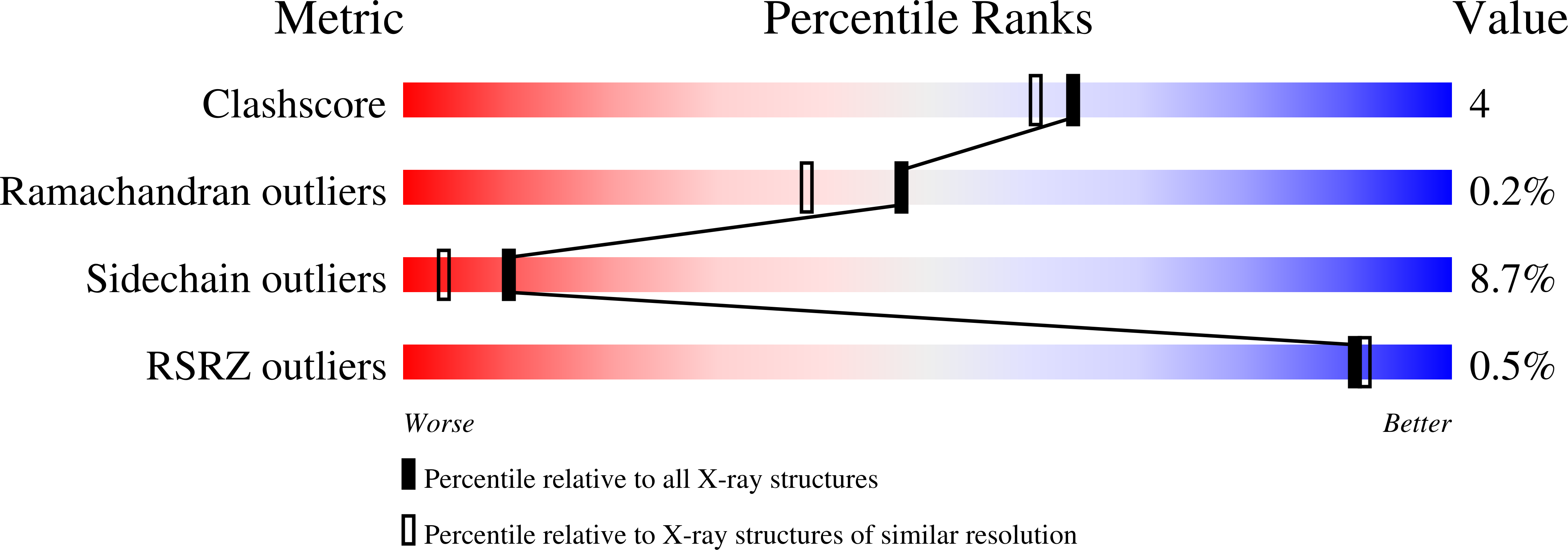
PDB ID:
1D7F
Keywords:
Title:
CRYSTAL STRUCTURE OF ASPARAGINE 233-REPLACED CYCLODEXTRIN GLUCANOTRANSFERASE FROM ALKALOPHILIC BACILLUS SP. 1011 DETERMINED AT 1.9 A RESOLUTION
Biological Source:
Source Organism(s):
Bacillus sp. (Taxon ID: 1410)
Expression System(s):
Method Details:
Experimental Method:
Resolution:
1.90 Å
R-Value Free:
0.20
R-Value Work:
0.15
R-Value Observed:
0.15
Space Group:
P 1