Deposition Date
1999-10-04
Release Date
2000-01-20
Last Version Date
2024-02-07
Entry Detail
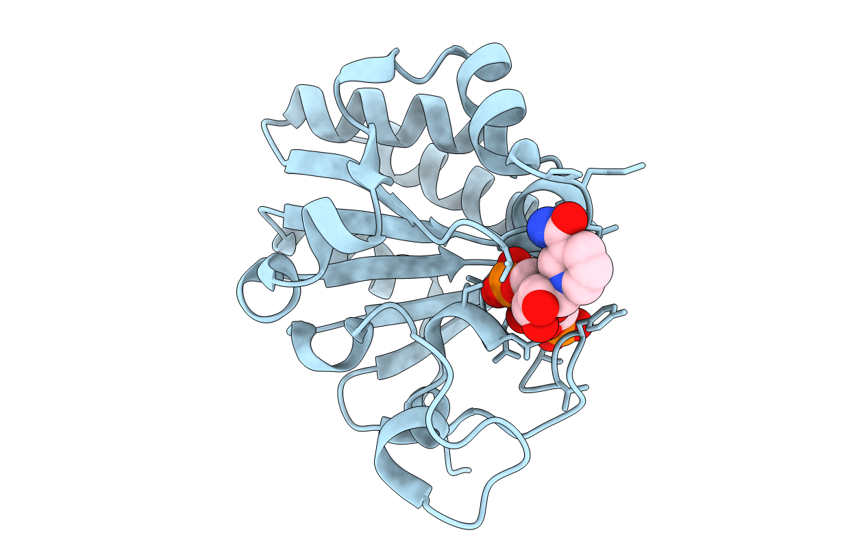
PDB ID:
1D4O
Keywords:
Title:
CRYSTAL STRUCTURE OF TRANSHYDROGENASE DOMAIN III AT 1.2 ANGSTROMS RESOLUTION
Biological Source:
Source Organism(s):
Bos taurus (Taxon ID: 9913)
Expression System(s):
Method Details:
Experimental Method:
Resolution:
1.21 Å
R-Value Free:
0.22
R-Value Work:
0.16
R-Value Observed:
0.16
Space Group:
P 1