1D3G image
Deposition Date 1999-09-29
Release Date 2000-09-13
Last Version Date 2024-02-07
Entry Detail
PDB ID:
1D3G
Keywords:
Title:
HUMAN DIHYDROOROTATE DEHYDROGENASE COMPLEXED WITH BREQUINAR ANALOG
Biological Source:
Source Organism:
Homo sapiens (Taxon ID: 9606)
Host Organism:
Method Details:
Experimental Method:
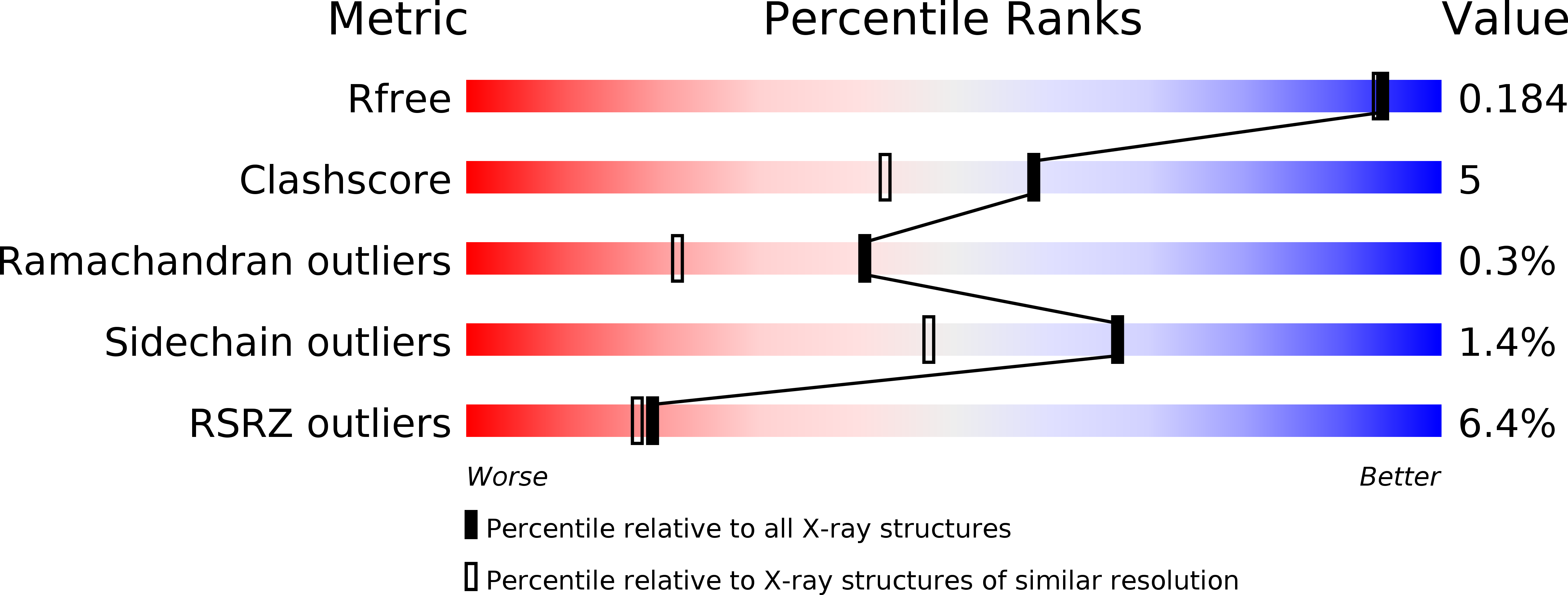
Resolution:
1.60 Å
R-Value Free:
0.18
R-Value Work:
0.16
Space Group:
P 32 2 1
Macromolecular Entities
Polymer Type:polypeptide(L)
Molecule:DIHYDROOROTATE DEHYDROGENASE
Gene (Uniprot):DHODH
Chain IDs:A
Chain Length:367
Number of Molecules:1
Biological Source:Homo sapiens
Primary Citation
Structures of human dihydroorotate dehydrogenase in complex with antiproliferative agents.
Structure Fold.Des. 8 25 33 (2000)
PMID: 10673429 DOI: 10.1016/S0969-2126(00)00077-0

Abstact

BACKGROUND: Dihydroorotate dehydrogenase (DHODH) catalyzes the fourth committed step in the de novo biosynthesis of pyrimidines. As rapidly proliferating human T cells have an exceptional requirement for de novo pyrimidine biosynthesis, small molecule DHODH inhibitors constitute an attractive therapeutic approach to autoimmune diseases, immunosuppression, and cancer. Neither the structure of human DHODH nor any member of its family was known. RESULTS: The high-resolution crystal structures of human DHODH in complex with two different inhibitors have been solved. The initial set of phases was obtained using multiwavelength anomalous diffraction phasing with selenomethionine-containing DHODH. The structures have been refined to crystallographic R factors of 16.8% and 16.2% at resolutions of 1. 6 A and 1.8 A for inhibitors related to brequinar and leflunomide, respectively. CONCLUSIONS: Human DHODH has two domains: an alpha/beta-barrel domain containing the active site and an alpha-helical domain that forms the opening of a tunnel leading to the active site. Both inhibitors share a common binding site in this tunnel, and differences in the binding region govern drug sensitivity or resistance. The active site of human DHODH is generally similar to that of the previously reported bacterial active site. The greatest differences are that the catalytic base removing the proton from dihydroorotate is a serine rather than a cysteine, and that packing of the flavin mononucleotide in its binding site is tighter.

Legend

Protein

Chemical

Disease

Primary Citation of related structures