1D0N image
Deposition Date 1999-09-13
Release Date 1999-09-15
Last Version Date 2024-10-30
Entry Detail
PDB ID:
1D0N
Title:
THE CRYSTAL STRUCTURE OF CALCIUM-FREE EQUINE PLASMA GELSOLIN.
Biological Source:
Source Organism:
Equus caballus (Taxon ID: 9796)
Method Details:
Experimental Method:
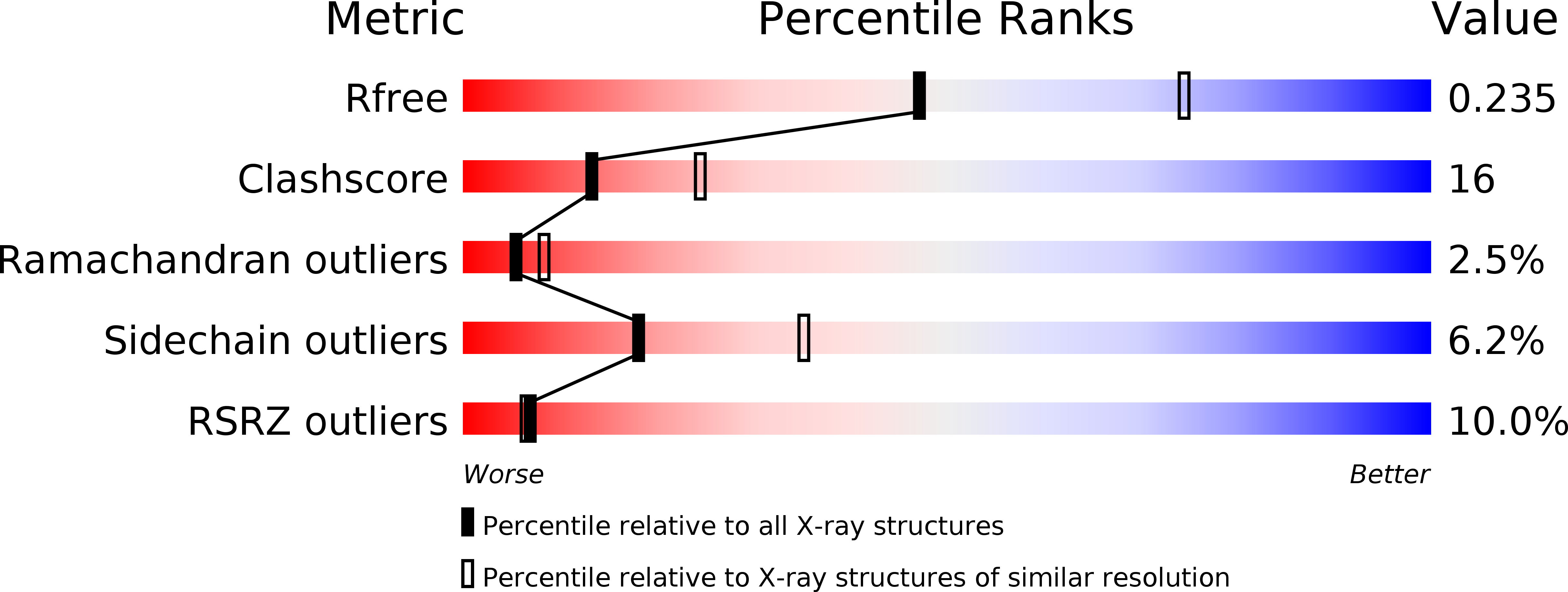
Resolution:
2.50 Å
R-Value Free:
0.23
R-Value Work:
0.20
R-Value Observed:
0.21
Space Group:
P 4 21 2
Macromolecular Entities
Structures with similar UniProt ID
Protein Blast
Polymer Type:polypeptide(L)
Molecule:HORSE PLASMA GELSOLIN
Gene (Uniprot):GSN
Chain IDs:A, B
Chain Length:729
Number of Molecules:2
Biological Source:Equus caballus
Primary Citation
The crystal structure of plasma gelsolin: implications for actin severing, capping, and nucleation.
Cell(Cambridge,Mass.) 90 661 670 (1997)
PMID: 9288746 DOI: 10.1016/S0092-8674(00)80527-9

Abstact

The structure of gelsolin has been determined by crystallography and comprises six structurally related domains that, in a Ca2+-free environment, pack together to form a compact globular structure in which the putative actin-binding sequences are not sufficiently exposed to enable binding to occur. We propose that binding Ca2+ can release the connections that join the N- and C-terminal halves of gelsolin, enabling each half to bind actin relatively independently. Domain shifts are proposed in response to Ca2+ as bases for models of how gelsolin acts to sever, cap, or nucleate F-actin filaments. The structure also invites discussion of polyphosphoinositide binding to segment 2 and suggests how mutation at Asp-187 could initiate a series of events that lead to deposition of amyloid plaques, as observed in victims of familial amyloidosis (Finnish type).

Legend

Protein

Chemical

Disease

Primary Citation of related structures
Feedback Form
Name
Email
Institute
Feedback