Deposition Date
1999-09-09
Release Date
2000-09-09
Last Version Date
2024-10-16
Entry Detail
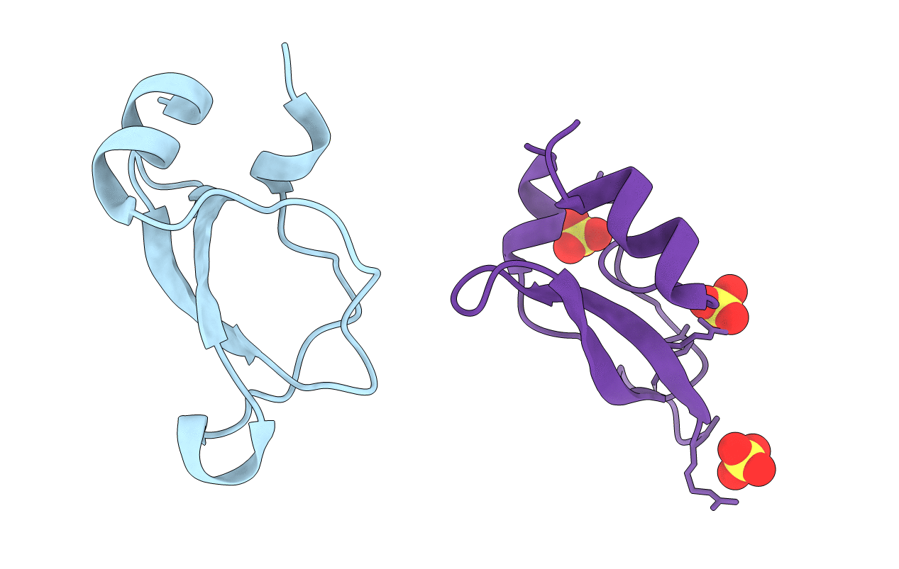
PDB ID:
1D0D
Keywords:
Title:
CRYSTAL STRUCTURE OF TICK ANTICOAGULANT PROTEIN COMPLEXED WITH BOVINE PANCREATIC TRYPSIN INHIBITOR
Biological Source:
Source Organism(s):
Ornithodoros moubata (Taxon ID: 6938)
Bos taurus (Taxon ID: 9913)
Bos taurus (Taxon ID: 9913)
Method Details:
Experimental Method:
Resolution:
1.62 Å
R-Value Free:
0.21
R-Value Work:
0.18
R-Value Observed:
0.19
Space Group:
P 41