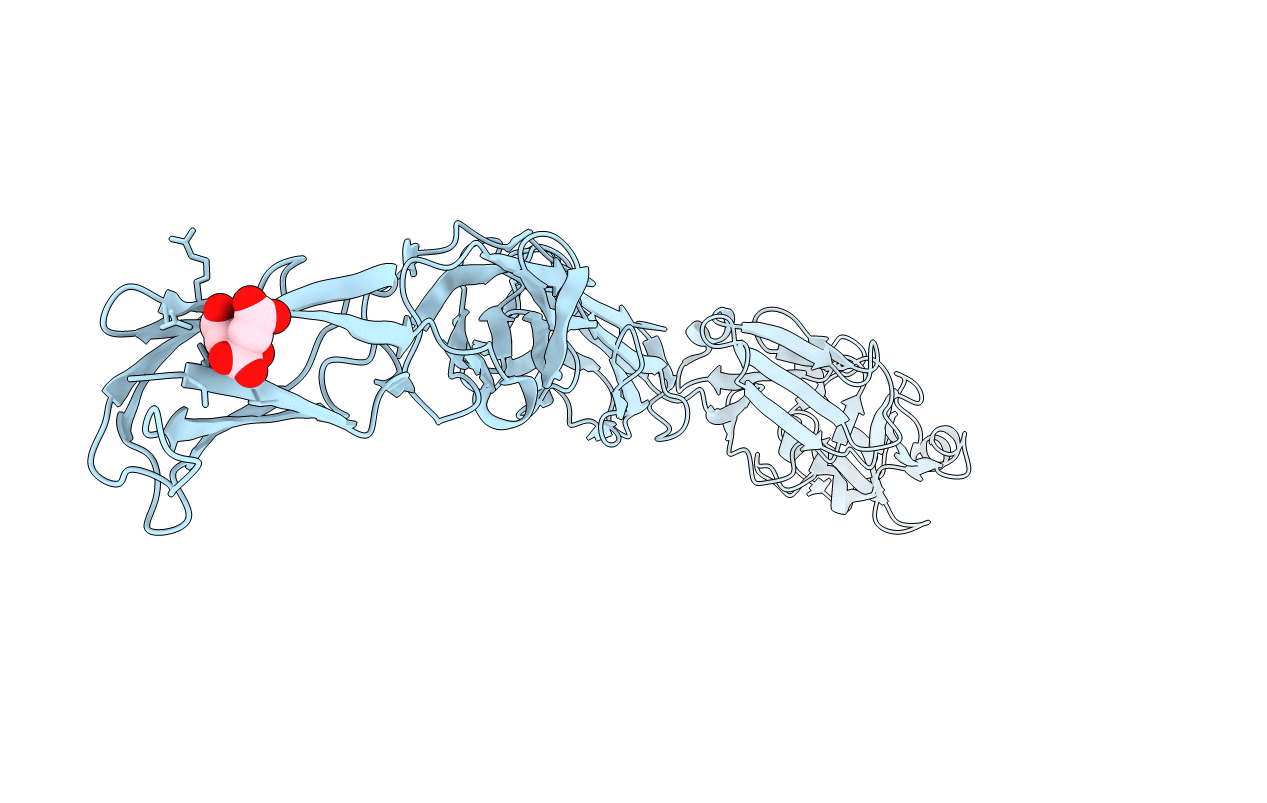
1CWV image
Deposition Date 1999-08-26
Release Date 1999-10-13
Last Version Date 2024-11-13
Entry Detail
PDB ID:
1CWV
Title:
CRYSTAL STRUCTURE OF INVASIN: A BACTERIAL INTEGRIN-BINDING PROTEIN
Biological Source:
Source Organism:
Host Organism:
Method Details:
Experimental Method:
Resolution:
2.30 Å
R-Value Free:
0.27
R-Value Work:
0.22
R-Value Observed:
0.22
Space Group:
P 1 21 1
Macromolecular Entities
Structures with similar UniProt ID
Protein Blast
Polymer Type:polypeptide(L)
Molecule:INVASIN
Gene (Uniprot):YPTB1668
Chain IDs:A
Chain Length:492
Number of Molecules:1
Biological Source:Yersinia pseudotuberculosis
Ligand Molecules
Primary Citation
Crystal structure of invasin: a bacterial integrin-binding protein.
Science 286 291 295 (1999)
PMID: 10514372 DOI: 10.1126/science.286.5438.291

Abstact

The Yersinia pseudotuberculosis invasin protein promotes bacterial entry by binding to host cell integrins with higher affinity than natural substrates such as fibronectin. The 2.3 angstrom crystal structure of the invasin extracellular region reveals five domains that form a 180 angstrom rod with structural similarities to tandem fibronectin type III domains. The integrin-binding surfaces of invasin and fibronectin include similarly located key residues, but in the context of different folds and surface shapes. The structures of invasin and fibronectin provide an example of convergent evolution, in which invasin presents an optimized surface for integrin binding, in comparison with host substrates.

Legend

Protein

Chemical

Disease

Primary Citation of related structures