Deposition Date
1998-06-05
Release Date
1998-08-12
Last Version Date
2023-08-09
Entry Detail
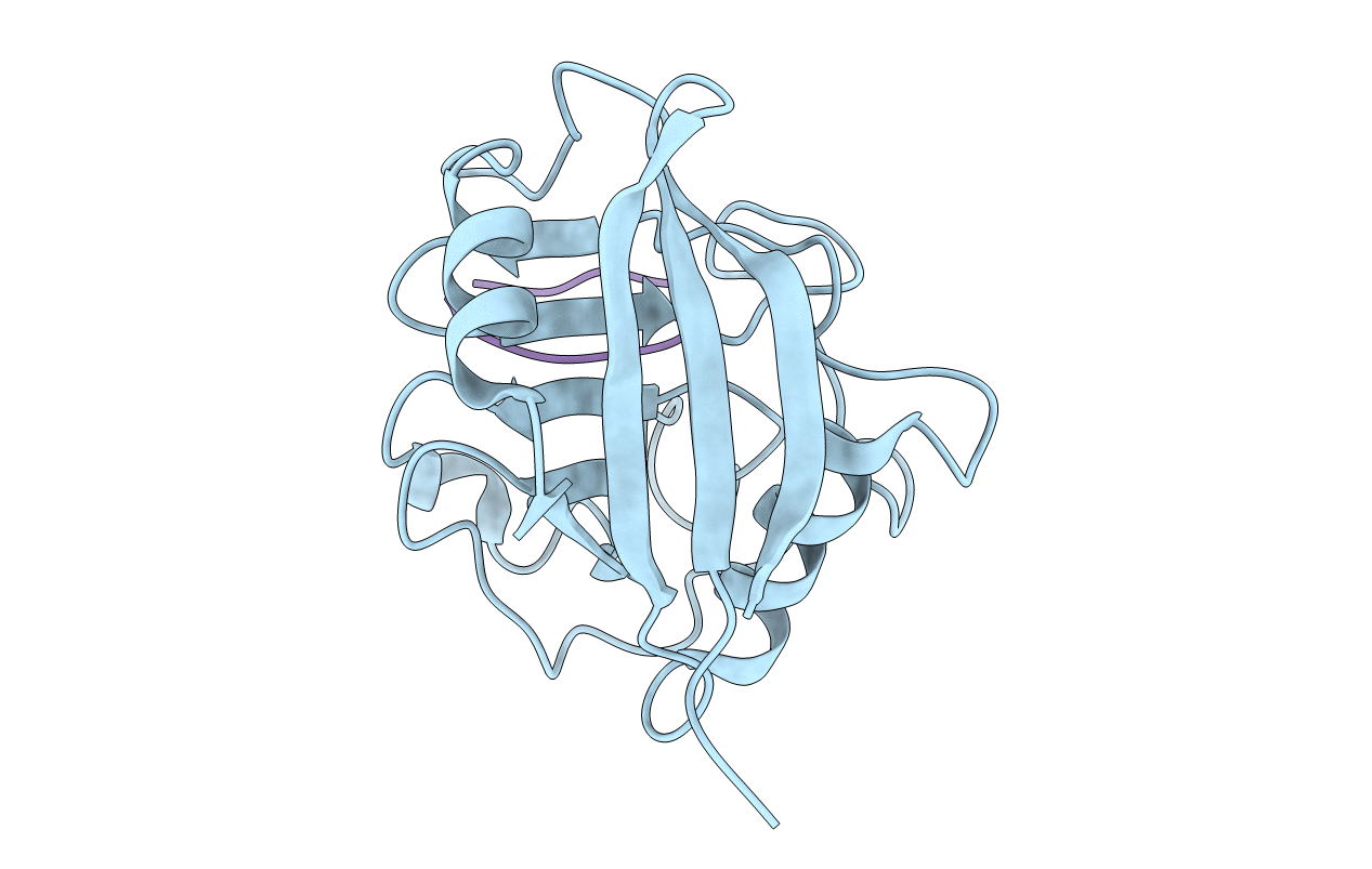
PDB ID:
1CWO
Keywords:
Title:
HUMAN CYCLOPHILIN A COMPLEXED WITH THR2, LEU5, D-HIV8, LEU10 CYCLOSPORIN
Biological Source:
Source Organism(s):
HOMO SAPIENS (Taxon ID: 9606)
TOLYPOCLADIUM INFLATUM (Taxon ID: 29910)
TOLYPOCLADIUM INFLATUM (Taxon ID: 29910)
Expression System(s):
Method Details: