Deposition Date
1999-08-25
Release Date
1999-09-01
Last Version Date
2024-02-07
Entry Detail
PDB ID:
1CW1
Keywords:
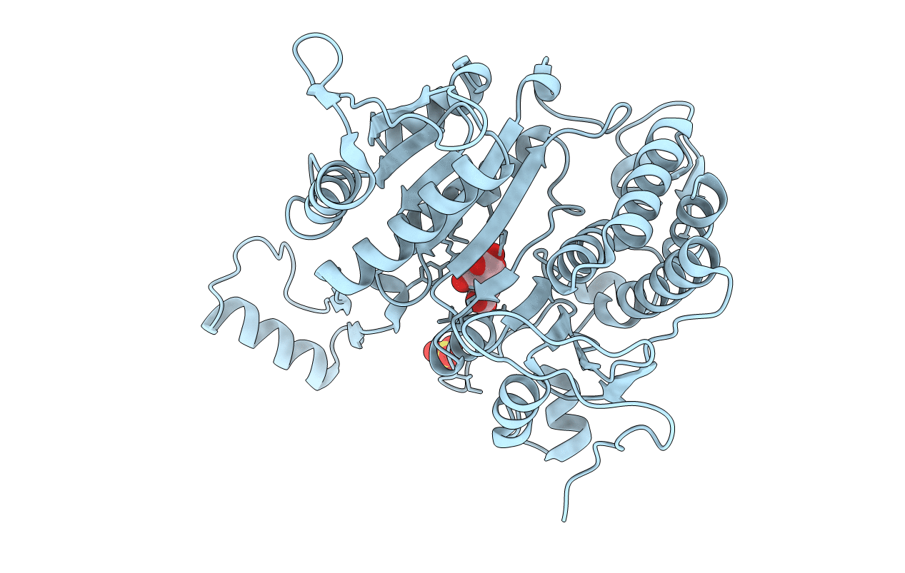
Title:
CRYSTAL STRUCTURE OF ISOCITRATE DEHYDROGENASE MUTANT K230M BOUND TO ISOCITRATE AND MN2+
Biological Source:
Source Organism(s):
Escherichia coli (Taxon ID: 562)
Expression System(s):
Method Details:
Experimental Method:
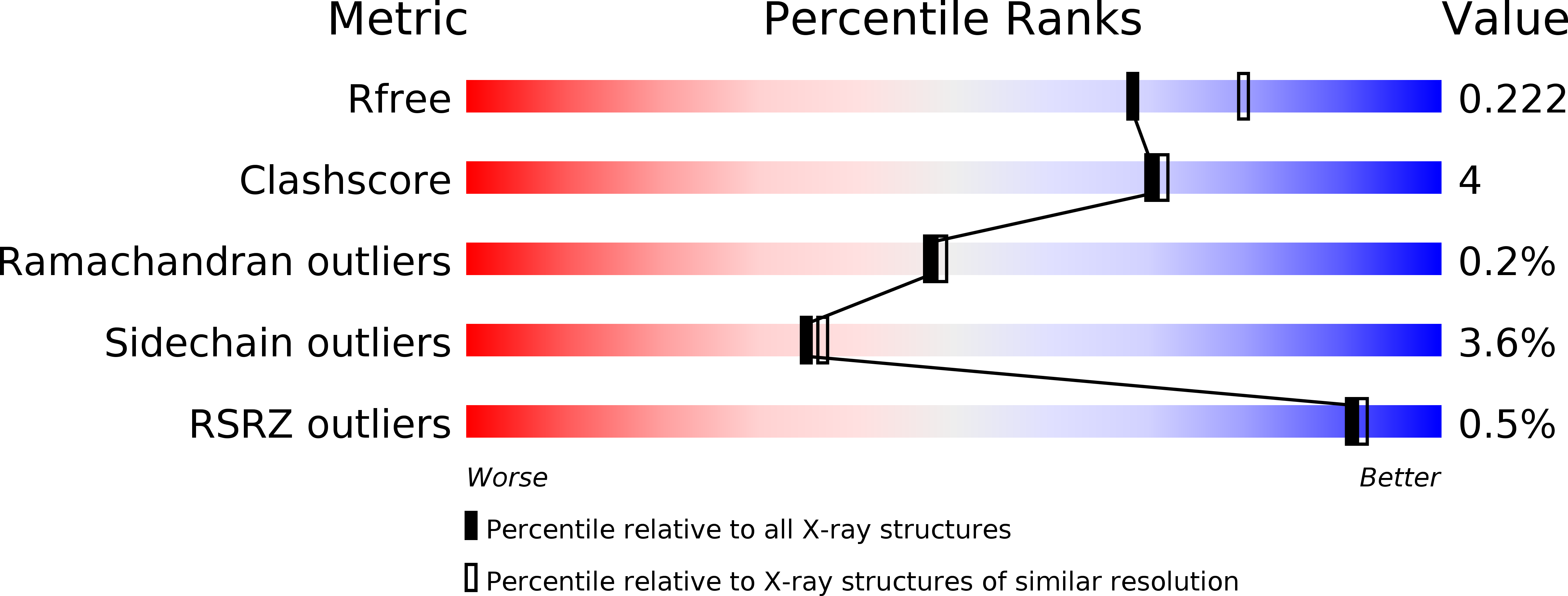
Resolution:
2.10 Å
R-Value Free:
0.23
R-Value Work:
0.19
R-Value Observed:
0.19
Space Group:
P 43 21 2