1CW0 image
Deposition Date 1999-08-25
Release Date 1999-12-12
Last Version Date 2023-08-09
Entry Detail
PDB ID:
1CW0
Keywords:
Title:
CRYSTAL STRUCTURE ANALYSIS OF VERY SHORT PATCH REPAIR (VSR) ENDONUCLEASE IN COMPLEX WITH A DUPLEX DNA
Biological Source:
Source Organism:
Escherichia coli (Taxon ID: 562)
Host Organism:
Method Details:
Experimental Method:
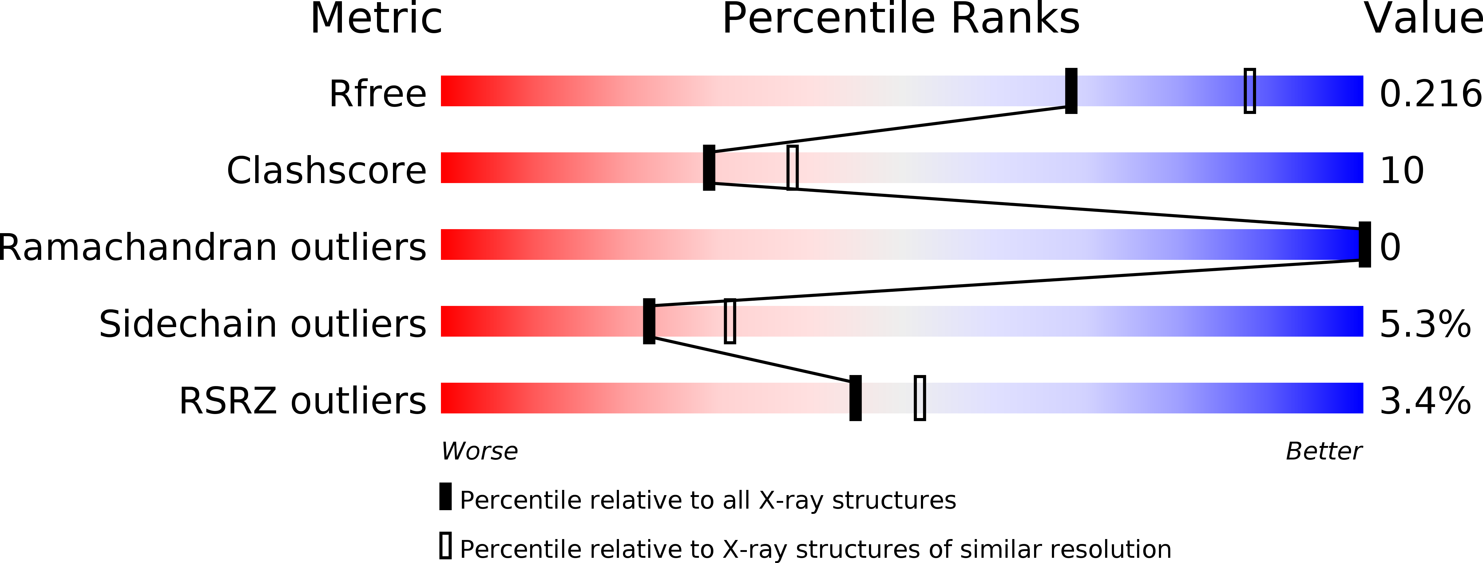
Resolution:
2.30 Å
R-Value Free:
0.23
R-Value Work:
0.20
R-Value Observed:
0.20
Space Group:
P 65
Macromolecular Entities
Structures with similar UniProt ID
Protein Blast
Polymer Type:polypeptide(L)
Molecule:PROTEIN (DNA MISMATCH ENDONUCLEASE)
Gene (Uniprot):vsr
Chain IDs:D (auth: A)
Chain Length:155
Number of Molecules:1
Biological Source:Escherichia coli
Primary Citation
Recognition of a TG mismatch: the crystal structure of very short patch repair endonuclease in complex with a DNA duplex.
Cell(Cambridge,Mass.) 99 615 623 (1999)
PMID: 10612397 DOI: 10.1016/S0092-8674(00)81550-0

Abstact

The crystal structure of very short patch repair (Vsr) endonuclease, in complex with Mg2+ and with duplex DNA containing a TG mismatch, has been determined at 2.3 A resolution. In E. coli, the enzyme recognizes a TG mismatched base pair, generated after spontaneous deamination of methylated cytosines, and cleaves the phosphate backbone on the 5' side of the thymine. Extensive interactions between the DNA and the protein characterize a novel recognition mechanism, where three aromatic residues intercalate from the major groove into the DNA to strikingly deform the base pair stacking. With the presence of a cleaved DNA intermediate in the active center, the structure of the Vsr/DNA complex provides detailed insights into the catalytic mechanism for endonuclease activity.

Legend

Protein

Chemical

Disease

Primary Citation of related structures