Deposition Date
1999-08-24
Release Date
1999-08-31
Last Version Date
2024-11-13
Entry Detail
PDB ID:
1CVW
Keywords:
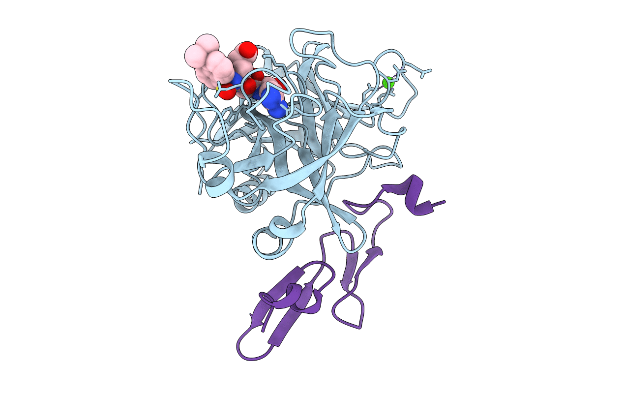
Title:
Crystal structure of active site-inhibited human coagulation factor VIIA (DES-GLA)
Biological Source:
Source Organism(s):
Homo sapiens (Taxon ID: 9606)
Expression System(s):
Method Details:
Experimental Method:
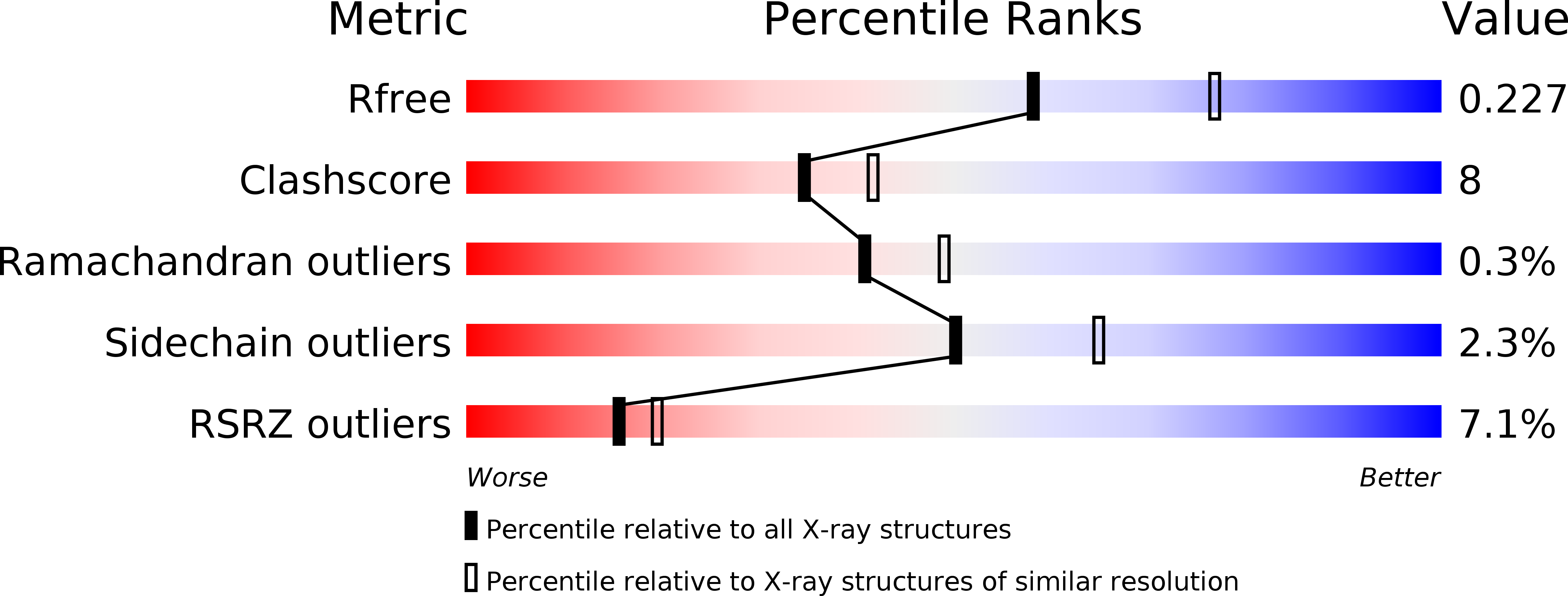
Resolution:
2.28 Å
R-Value Free:
0.23
R-Value Work:
0.20
R-Value Observed:
0.20
Space Group:
P 41 21 2